 Movie
Movie Controller


+ Open data

- Basic information
| Entry | Database: PDB / ID: 1g8w | ||||||
|---|---|---|---|---|---|---|---|
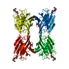
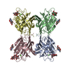
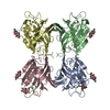
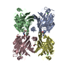
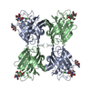
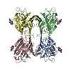
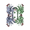
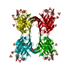
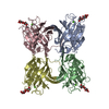
| Title | IMPROVED STRUCTURE OF PHYTOHEMAGGLUTININ-L FROM THE KIDNEY BEAN | ||||||
 Components Components | LEUCOAGGLUTINATING PHYTOHEMAGGLUTININ | ||||||
 Keywords Keywords | SUGAR BINDING PROTEIN / JELLY-ROLL FOLD / LEGUME LECTIN | ||||||
| Function / homology |  Function and homology information Function and homology information | ||||||
| Biological species |  Phaseolus vulgaris (common bean) Phaseolus vulgaris (common bean) | ||||||
| Method |  X-RAY DIFFRACTION / MOLECULAR REPLACEMENT / Resolution: 2.8 Å X-RAY DIFFRACTION / MOLECULAR REPLACEMENT / Resolution: 2.8 Å | ||||||
 Authors Authors | Buts, L. / Hamelryck, T.W. / Dao-Thi, M. / Loris, R. / Wyns, L. / Etzler, M.E. | ||||||
 Citation Citation | Journal: J.Mol.Biol. / Year: 2001 Title: Weak protein-protein interactions in lectins: the crystal structure of a vegetative lectin from the legume Dolichos biflorus. Authors: Buts, L. / Dao-Thi, M.H. / Loris, R. / Wyns, L. / Etzler, M. / Hamelryck, T. #1: Journal: J.Biol.Chem. / Year: 1996Title: The crystallographic structure of phytohemagglutinin-L Authors: Hamelryck, T.W. / Dao-Thi, M. / Poortmans, F. / Chrispeels, M.J. / Wyns, L. / Loris, R. #2: Journal: Proteins / Year: 1996Title: Crystallisation of glycosylated and non-glycosylated phytohemagglutinin-L Authors: Dao-Thi, M. / Hamelryck, T.W. / Poortmans, F. / Voelker, T.A. / Chrispeels, M.J. / Wyns, L. | ||||||
| History |
|
- Structure visualization
| Structure viewer | Molecule: MolmilJmol/JSmol |
|---|
- Downloads & links
-Download
| PDBx/mmCIF format | 1g8w.cif.gz | 187.7 KB | Display | PDBx/mmCIF format |
|---|---|---|---|---|
| PDB format | pdb1g8w.ent.gz | 150.2 KB | Display | PDB format |
| PDBx/mmJSON format | 1g8w.json.gz | Tree view | PDBx/mmJSON format | |
| Others |  Other downloads Other downloads |
-Validation report
| Arichive directory | https://data.pdbj.org/pub/pdb/validation_reports/g8/1g8wftp://data.pdbj.org/pub/pdb/validation_reports/g8/1g8w | HTTPS FTP |
|---|
-Related structure data
| Related structure data | 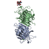 1g7yC 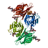 1g9fC 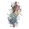 1lesS S: Starting model for refinement C: citing same article ( |
|---|---|
| Similar structure data |

-Links
 PDBj
PDBj

- Assembly
| Deposited unit | 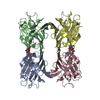
| ||||||||
|---|---|---|---|---|---|---|---|---|---|
| 1 |
| ||||||||
| Unit cell |
|
-Components
| #1: Protein | Mass: 25397.959 Da / Num. of mol.: 4 / Fragment: LEUCOAGGLUTINATING FRACTION OF THE SEED LECTIN / Source method: isolated from a natural source / Source: (natural) Phaseolus vulgaris (common bean) / Organ: SEED / References: UniProt: P05087#2: Sugar | ChemComp-NAG /   Type: D-saccharide, beta linking / Mass: 221.208 Da / Num. of mol.: 4 Type: D-saccharide, beta linking / Mass: 221.208 Da / Num. of mol.: 4Source method: isolated from a genetically manipulated source Formula: C8H15NO6 #3: Chemical | ChemComp-MN /   Mass: 54.938 Da / Num. of mol.: 4 / Source method: obtained synthetically / Formula: Mn Mass: 54.938 Da / Num. of mol.: 4 / Source method: obtained synthetically / Formula: Mn#4: Chemical | ChemComp-CA /   Mass: 40.078 Da / Num. of mol.: 4 / Source method: obtained synthetically / Formula: Ca Mass: 40.078 Da / Num. of mol.: 4 / Source method: obtained synthetically / Formula: Ca#5: Water | ChemComp-HOH / |  Mass: 18.015 Da / Num. of mol.: 16 / Source method: isolated from a natural source / Formula: H2O Mass: 18.015 Da / Num. of mol.: 16 / Source method: isolated from a natural source / Formula: H2OHas protein modification | Y | |
|---|
-Experimental details
-Experiment
| Experiment | Method: X-RAY DIFFRACTION / Number of used crystals: 1 |
|---|
- Sample preparation
| Crystal | Density Matthews: 2.85 Å3/Da / Density % sol: 56.89 % | ||||||||||||||||||||||||||||||
|---|---|---|---|---|---|---|---|---|---|---|---|---|---|---|---|---|---|---|---|---|---|---|---|---|---|---|---|---|---|---|---|
| Crystal grow | Temperature: 293 K / Method: vapor diffusion, hanging drop / pH: 8.5 Details: 5 mg/ml protein; 100 mM TRIS/HCl; 8% (w/v) PEG(6000), pH 8.5, VAPOR DIFFUSION, HANGING DROP, temperature 293K | ||||||||||||||||||||||||||||||
| Crystal grow | *PLUS pH: 5.6 | ||||||||||||||||||||||||||||||
| Components of the solutions | *PLUS
|
-Data collection
| Diffraction | Mean temperature: 298 K |
|---|---|
| Diffraction source | Source: ROTATING ANODE / Type: RIGAKU / Wavelength: 1.5418 Å |
| Detector | Type: ENRAF-NONIUS FAST / Detector: DIFFRACTOMETER / Date: Aug 1, 1995 |
| Radiation | Monochromator: Ni FILTER / Protocol: SINGLE WAVELENGTH / Monochromatic (M) / Laue (L): M / Scattering type: x-ray |
| Radiation wavelength | Wavelength: 1.5418 Å / Relative weight: 1 |
| Reflection | Resolution: 2.8→10 Å / Num. all: 105939 / Num. obs: 100006 / % possible obs: 94.4 % / Observed criterion σ(F): 0 / Observed criterion σ(I): 5 / Redundancy: 3.8 % / Biso Wilson estimate: 72 Å2 / Rmerge(I) obs: 0.107 / Net I/σ(I): 11.23 |
| Reflection shell | Resolution: 2.8→2.95 Å / Mean I/σ(I) obs: 3.2 / % possible all: 82.1 |
| Reflection | *PLUS Lowest resolution: 20 Å / Num. all: 100006 / Num. obs: 26546 / Num. measured all: 105939 |
| Reflection shell | *PLUS % possible obs: 82.1 % |
- Processing
| Software |
| ||||||||||||||||||||||||||||||||||||||||
|---|---|---|---|---|---|---|---|---|---|---|---|---|---|---|---|---|---|---|---|---|---|---|---|---|---|---|---|---|---|---|---|---|---|---|---|---|---|---|---|---|---|
| Refinement | Method to determine structure: MOLECULAR REPLACEMENT Starting model: PDB ENTRY 1LES (lentil lectin) Resolution: 2.8→20 Å / Rfactor Rfree error: 0.004 / Data cutoff high absF: 3547881.72 / Data cutoff low absF: 0 / Isotropic thermal model: RESTRAINED / Cross valid method: THROUGHOUT / σ(F): 0 / σ(I): 0 / Stereochemistry target values: Engh & Huber
| ||||||||||||||||||||||||||||||||||||||||
| Solvent computation | Solvent model: FLAT MODEL / Bsol: 19.88 Å2 / ksol: 0.262 e/Å3 | ||||||||||||||||||||||||||||||||||||||||
| Displacement parameters | Biso mean: 34.3 Å2
| ||||||||||||||||||||||||||||||||||||||||
| Refine analyze |
| ||||||||||||||||||||||||||||||||||||||||
| Refinement step | Cycle: LAST / Resolution: 2.8→20 Å
| ||||||||||||||||||||||||||||||||||||||||
| Refine LS restraints |
| ||||||||||||||||||||||||||||||||||||||||
| Refine LS restraints NCS | NCS model details: CONSTR | ||||||||||||||||||||||||||||||||||||||||
| LS refinement shell | Resolution: 2.8→2.97 Å / Rfactor Rfree error: 0.019 / Total num. of bins used: 6
| ||||||||||||||||||||||||||||||||||||||||
| Software | *PLUS Name: CNS / Version: 1 / Classification: refinement | ||||||||||||||||||||||||||||||||||||||||
| Refinement | *PLUS Highest resolution: 2.8 Å / Lowest resolution: 20 Å / σ(F): 0 / % reflection Rfree: 9.9 % | ||||||||||||||||||||||||||||||||||||||||
| Solvent computation | *PLUS | ||||||||||||||||||||||||||||||||||||||||
| Displacement parameters | *PLUS Biso mean: 34.3 Å2 | ||||||||||||||||||||||||||||||||||||||||
| Refine LS restraints | *PLUS
| ||||||||||||||||||||||||||||||||||||||||
| LS refinement shell | *PLUS Rfactor Rfree: 0.31 / % reflection Rfree: 9.9 % / Rfactor Rwork: 0.28 |
