 Movie
Movie Controller


+ Open data

- Basic information
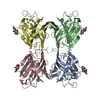
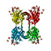
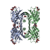
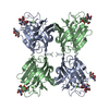
| Entry | Database: PDB / ID: 1sbe | |||||||||
|---|---|---|---|---|---|---|---|---|---|---|
| Title | SOYBEAN AGGLUTININ FROM GLYCINE MAX | |||||||||
 Components Components | SOYBEAN AGGLUTININ | |||||||||
 Keywords Keywords | LECTIN / AGGLUTININ | |||||||||
| Function / homology |  Function and homology information Function and homology information | |||||||||
| Biological species |  | |||||||||
| Method |  X-RAY DIFFRACTION / MOLECULAR DYNAMICS VS 1SBA / Resolution: 2.8 Å X-RAY DIFFRACTION / MOLECULAR DYNAMICS VS 1SBA / Resolution: 2.8 Å | |||||||||
 Authors Authors | Dessen, A. / Olsen, L.R. / Gupta, D. / Sabesan, S. / Sacchettini, J.C. / Brewer, C.F. | |||||||||
 Citation Citation | Journal: Biochemistry / Year: 1997 Title: X-ray crystallographic studies of unique cross-linked lattices between four isomeric biantennary oligosaccharides and soybean agglutinin. Authors: Olsen, L.R. / Dessen, A. / Gupta, D. / Sabesan, S. / Sacchettini, J.C. / Brewer, C.F. #1: Journal: Biochemistry / Year: 1995Title: X-Ray Crystal Structure of the Soybean Agglutinin Cross-Linked with a Biantennary Analog of the Blood Group I Carbohydrate Antigen Authors: Dessen, A. / Gupta, D. / Sabesan, S. / Brewer, C.F. / Sacchettini, J.C. | |||||||||
| History |
|
- Structure visualization
| Structure viewer | Molecule: MolmilJmol/JSmol |
|---|
- Downloads & links
-Download
| PDBx/mmCIF format | 1sbe.cif.gz | 60.3 KB | Display | PDBx/mmCIF format |
|---|---|---|---|---|
| PDB format | pdb1sbe.ent.gz | 41.4 KB | Display | PDB format |
| PDBx/mmJSON format | 1sbe.json.gz | Tree view | PDBx/mmJSON format | |
| Others |  Other downloads Other downloads |
-Validation report
| Arichive directory | https://data.pdbj.org/pub/pdb/validation_reports/sb/1sbeftp://data.pdbj.org/pub/pdb/validation_reports/sb/1sbe | HTTPS FTP |
|---|
-Related structure data


-Links
 PDBj
PDBj

- Assembly
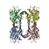
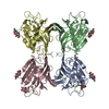
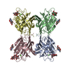
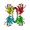
| Deposited unit | 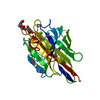
| ||||||||
|---|---|---|---|---|---|---|---|---|---|
| 1 | 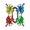
| ||||||||
| Unit cell |
|
-Components
-Protein , 1 types, 1 molecules A
| #1: Protein | Mass: 27595.816 Da / Num. of mol.: 1 / Source method: isolated from a natural source / Source: (natural) |
|---|
-Sugars , 2 types, 2 molecules 
| #2: Polysaccharide | beta-D-galactopyranose-(1-4)-2-acetamido-2-deoxy-beta-D-glucopyranose Source method: isolated from a genetically manipulated source |
|---|---|
| #3: Sugar | ChemComp-NAG /  Type: D-saccharide, beta linking / Mass: 221.208 Da / Num. of mol.: 1 Type: D-saccharide, beta linking / Mass: 221.208 Da / Num. of mol.: 1Source method: isolated from a genetically manipulated source Formula: C8H15NO6 |
-Non-polymers , 3 types, 22 molecules 


| #4: Chemical | ChemComp-MN /  Mass: 54.938 Da / Num. of mol.: 1 / Source method: obtained synthetically / Formula: Mn Mass: 54.938 Da / Num. of mol.: 1 / Source method: obtained synthetically / Formula: Mn |
|---|---|
| #5: Chemical | ChemComp-CA /  Mass: 40.078 Da / Num. of mol.: 1 / Source method: obtained synthetically / Formula: Ca Mass: 40.078 Da / Num. of mol.: 1 / Source method: obtained synthetically / Formula: Ca |
| #6: Water | ChemComp-HOH / Mass: 18.015 Da / Num. of mol.: 20 / Source method: isolated from a natural source / Formula: H2O |
-Details
| Compound details | RATHER THAN PROTEIN-PROTEIN CONTACTS, THE DOMINANT FORCE IN THESE CRYSTALS IS SACCHARIDE-PROTEIN ...RATHER THAN PROTEIN-PROTEIN CONTACTS, THE DOMINANT FORCE IN THESE CRYSTALS IS SACCHARIDE |
|---|---|
| Has protein modification | Y |
| Nonpolymer details | THESE COORDINATE |
-Experimental details
-Experiment
| Experiment | Method: X-RAY DIFFRACTION / Number of used crystals: 1 |
|---|
- Sample preparation
| Crystal | Density Matthews: 5.7 Å3/Da / Density % sol: 78 % | ||||||||||||||||||||||||||||||||||||||||||||||||||||||||||||||||||
|---|---|---|---|---|---|---|---|---|---|---|---|---|---|---|---|---|---|---|---|---|---|---|---|---|---|---|---|---|---|---|---|---|---|---|---|---|---|---|---|---|---|---|---|---|---|---|---|---|---|---|---|---|---|---|---|---|---|---|---|---|---|---|---|---|---|---|---|
| Crystal grow | pH: 7.2 Details: PROTEIN WAS CRYSTALLIZED FROM 0.1 M HEPES, PH 7.2 1MM CACL2, 1MM MNCL2 AND 0.15 M NACL | ||||||||||||||||||||||||||||||||||||||||||||||||||||||||||||||||||
| Crystal | *PLUS Density % sol: 70 % | ||||||||||||||||||||||||||||||||||||||||||||||||||||||||||||||||||
| Crystal grow | *PLUS Temperature: 4 ℃ / Method: vapor diffusion, hanging drop / Details: Dessen, A., (1995) Biochemistry, 34, 4933. | ||||||||||||||||||||||||||||||||||||||||||||||||||||||||||||||||||
| Components of the solutions | *PLUS
|
-Data collection
| Diffraction | Mean temperature: 293 K |
|---|---|
| Diffraction source | Source: ROTATING ANODE / Type: RIGAKU RUH2R / Wavelength: 1.5418 |
| Detector | Type: SIEMENS / Detector: AREA DETECTOR / Date: Dec 1, 1993 / Details: COLLIMATOR |
| Radiation | Monochromator: NI FILTER / Monochromatic (M) / Laue (L): M / Scattering type: x-ray |
| Radiation wavelength | Wavelength: 1.5418 Å / Relative weight: 1 |
| Reflection | Resolution: 2.8→99 Å / Num. obs: 16581 / % possible obs: 84 % / Observed criterion σ(I): 2 / Redundancy: 3.7 % / Biso Wilson estimate: 7.79 Å2 / Rmerge(I) obs: 0.139 / Rsym value: 0.139 / Net I/σ(I): 4.1 |
| Reflection shell | Resolution: 2.8→2.97 Å / Redundancy: 3 % / Rmerge(I) obs: 0.54 / Mean I/σ(I) obs: 0.6 / Rsym value: 0.54 / % possible all: 67 |
| Reflection | *PLUS Num. measured all: 61134 |
| Reflection shell | *PLUS % possible obs: 67 % |
- Processing
| Software |
| ||||||||||||||||||||||||||||||||||||||||||||||||||
|---|---|---|---|---|---|---|---|---|---|---|---|---|---|---|---|---|---|---|---|---|---|---|---|---|---|---|---|---|---|---|---|---|---|---|---|---|---|---|---|---|---|---|---|---|---|---|---|---|---|---|---|
| Refinement | Method to determine structure: MOLECULAR DYNAMICS VS 1SBA / Resolution: 2.8→99 Å / Isotropic thermal model: TNT BCORREL / σ(F): 0 / Stereochemistry target values: TNT PROTGEO Details: INITIAL REFINEMENTS USED SLOWCOOL ROUTINE FROM X-PLOR.
| ||||||||||||||||||||||||||||||||||||||||||||||||||
| Solvent computation | Bsol: 398 Å2 / ksol: 0.54 e/Å3 | ||||||||||||||||||||||||||||||||||||||||||||||||||
| Refinement step | Cycle: LAST / Resolution: 2.8→99 Å
| ||||||||||||||||||||||||||||||||||||||||||||||||||
| Refine LS restraints |
| ||||||||||||||||||||||||||||||||||||||||||||||||||
| Software | *PLUS Name: TNT / Version: 5E / Classification: refinement | ||||||||||||||||||||||||||||||||||||||||||||||||||
| Refinement | *PLUS Rfactor all: 0.212 | ||||||||||||||||||||||||||||||||||||||||||||||||||
| Solvent computation | *PLUS | ||||||||||||||||||||||||||||||||||||||||||||||||||
| Displacement parameters | *PLUS | ||||||||||||||||||||||||||||||||||||||||||||||||||
| Refine LS restraints | *PLUS
|
