 Movie
Movie Controller


+ Open data

- Basic information
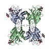
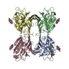
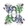
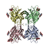
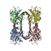
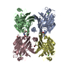
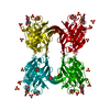
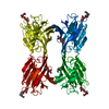
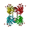
| Entry | Database: PDB / ID: 1qnw | ||||||
|---|---|---|---|---|---|---|---|
| Title | lectin II from Ulex europaeus | ||||||
 Components Components | CHITIN BINDING LECTIN, UEA-II | ||||||
 Keywords Keywords | LECTIN / CARBOHYDRATE BINDING | ||||||
| Function / homology |  Function and homology information Function and homology information | ||||||
| Biological species |  ULEX EUROPAEUS (furze) ULEX EUROPAEUS (furze) | ||||||
| Method |  X-RAY DIFFRACTION / SYNCHROTRON / MOLECULAR REPLACEMENT / Resolution: 2.35 Å X-RAY DIFFRACTION / SYNCHROTRON / MOLECULAR REPLACEMENT / Resolution: 2.35 Å | ||||||
 Authors Authors | Loris, R. / De Greve, H. / Dao-Thi, M.-H. / Messens, J. / Imberty, A. / Wyns, L. | ||||||
 Citation Citation | Journal: J.Mol.Biol. / Year: 2000 Title: Structural Basis of Carbohydrate Recognition by Lectin II from Ulex Europaeus, a Protein with a Promiscuous Carbohydrate Binding Site Authors: Loris, R. / De Greve, H. / Dao-Thi, M.-H. / Messens, J. / Imberty, A. / Wyns, L. #1: Journal: Curr.Opin.Struct.Biol. / Year: 1999 Title: Novel Structures of Plant Lectins and Their Complexes with Carbohydrates Authors: Bouckaert, J. / Hamelryck, T.W. / Wyns, L. / Loris, R. / Hamelryck, T.W. #2: Journal: Acta Crystallogr.,Sect.D / Year: 1998 Title: The Quaternary Structure of Uea-II, the Chitobiose Specific Lectin from Gorse Authors: Dao-Thi, M.-H. / Rizkallah, P. / Wyns, L. / Poortmans, F. / Loris, R. #3: Journal: Biochim.Biophys.Acta / Year: 1998 Title: Legume Lectin Structure Authors: Loris, R. / Hamelryck, T.W. / Bouckaert, J. / Wyns, L. | ||||||
| History |
|
- Structure visualization
| Structure viewer | Molecule: MolmilJmol/JSmol |
|---|
- Downloads & links
-Download
| PDBx/mmCIF format | 1qnw.cif.gz | 192.7 KB | Display | PDBx/mmCIF format |
|---|---|---|---|---|
| PDB format | pdb1qnw.ent.gz | 155.1 KB | Display | PDB format |
| PDBx/mmJSON format | 1qnw.json.gz | Tree view | PDBx/mmJSON format | |
| Others |  Other downloads Other downloads |
-Validation report
| Arichive directory | https://data.pdbj.org/pub/pdb/validation_reports/qn/1qnwftp://data.pdbj.org/pub/pdb/validation_reports/qn/1qnw | HTTPS FTP |
|---|
-Related structure data


-Links
 PDBj
PDBj

- Assembly
| Deposited unit | 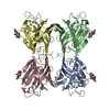
| ||||||||
|---|---|---|---|---|---|---|---|---|---|
| 1 |
| ||||||||
| Unit cell |
| ||||||||
| Components on special symmetry positions |
|
-Components
| #1: Protein | Mass: 26093.008 Da / Num. of mol.: 4 / Source method: isolated from a natural source / Source: (natural) ULEX EUROPAEUS (furze) / Organ: SEED / References: GenBank: AF190633, UniProt: P22973*PLUS#2: Chemical | ChemComp-MN /   Mass: 54.938 Da / Num. of mol.: 4 / Source method: obtained synthetically / Formula: Mn Mass: 54.938 Da / Num. of mol.: 4 / Source method: obtained synthetically / Formula: Mn#3: Chemical | ChemComp-CA /   Mass: 40.078 Da / Num. of mol.: 4 / Source method: obtained synthetically / Formula: Ca Mass: 40.078 Da / Num. of mol.: 4 / Source method: obtained synthetically / Formula: Ca#4: Sugar | ChemComp-NAG /   Type: D-saccharide, beta linking / Mass: 221.208 Da / Num. of mol.: 4 Type: D-saccharide, beta linking / Mass: 221.208 Da / Num. of mol.: 4Source method: isolated from a genetically manipulated source Formula: C8H15NO6 #5: Water | ChemComp-HOH / |  Mass: 18.015 Da / Num. of mol.: 201 / Source method: isolated from a natural source / Formula: H2O Mass: 18.015 Da / Num. of mol.: 201 / Source method: isolated from a natural source / Formula: H2OHas protein modification | Y | |
|---|
-Experimental details
-Experiment
| Experiment | Method: X-RAY DIFFRACTION / Number of used crystals: 2 |
|---|
- Sample preparation
| Crystal | Density Matthews: 2.67 Å3/Da / Density % sol: 53.94 % | ||||||||||||||||||||||||
|---|---|---|---|---|---|---|---|---|---|---|---|---|---|---|---|---|---|---|---|---|---|---|---|---|---|
| Crystal grow | pH: 6.5 / Details: pH 6.50 | ||||||||||||||||||||||||
| Crystal | *PLUS Density % sol: 45 % | ||||||||||||||||||||||||
| Crystal grow | *PLUS Method: vapor diffusion, hanging dropDetails: Dao-Thi M.H., (1998) Acta Crystallog. sect., D54, 844. | ||||||||||||||||||||||||
| Components of the solutions | *PLUS
|
-Data collection
| Diffraction | Mean temperature: 287 K |
|---|---|
| Diffraction source | Source: SYNCHROTRON / Site: SRS  / Beamline: PX9.6 / Wavelength: 0.87 / Beamline: PX9.6 / Wavelength: 0.87 |
| Detector | Type: MARRESEARCH / Detector: IMAGE PLATE / Details: MIRRORS |
| Radiation | Protocol: SINGLE WAVELENGTH / Monochromatic (M) / Laue (L): M / Scattering type: x-ray |
| Radiation wavelength | Wavelength: 0.87 Å / Relative weight: 1 |
| Reflection | Resolution: 2.35→50 Å / Num. obs: 45367 / % possible obs: 97 % / Observed criterion σ(I): -3 / Redundancy: 3.39 % / Rmerge(I) obs: 0.134 / Net I/σ(I): 9.99 |
| Reflection shell | Resolution: 2.35→2.45 Å / Redundancy: 1.66 % / Rmerge(I) obs: 0.542 / Mean I/σ(I) obs: 2.76 / % possible all: 94.6 |
| Reflection | *PLUS Num. measured all: 153615 |
| Reflection shell | *PLUS % possible obs: 94.6 % / Num. unique obs: 4365 / Num. measured obs: 7251 |
- Processing
| Software |
| ||||||||||||||||||||||||||||||||||||||||||||||||||||||||||||
|---|---|---|---|---|---|---|---|---|---|---|---|---|---|---|---|---|---|---|---|---|---|---|---|---|---|---|---|---|---|---|---|---|---|---|---|---|---|---|---|---|---|---|---|---|---|---|---|---|---|---|---|---|---|---|---|---|---|---|---|---|---|
| Refinement | Method to determine structure: MOLECULAR REPLACEMENT Starting model: PHA-L AND MODEL FROM SWISS-MODEL SERVER Resolution: 2.35→50 Å / Data cutoff high absF: 0 / Data cutoff low absF: 0 / Isotropic thermal model: RESTRAINED / Cross valid method: THROUGHOUT / σ(F): 0
| ||||||||||||||||||||||||||||||||||||||||||||||||||||||||||||
| Refinement step | Cycle: LAST / Resolution: 2.35→50 Å
| ||||||||||||||||||||||||||||||||||||||||||||||||||||||||||||
| Refine LS restraints |
| ||||||||||||||||||||||||||||||||||||||||||||||||||||||||||||
| LS refinement shell | Resolution: 2.35→2.46 Å / Total num. of bins used: 8
| ||||||||||||||||||||||||||||||||||||||||||||||||||||||||||||
| Software | *PLUS Name: X-PLOR / Version: 3.8 / Classification: refinement | ||||||||||||||||||||||||||||||||||||||||||||||||||||||||||||
| Refine LS restraints | *PLUS
| ||||||||||||||||||||||||||||||||||||||||||||||||||||||||||||
| LS refinement shell | *PLUS Rfactor obs: 0.281 |
