 Movie
Movie Controller


+ Open data

- Basic information
| Entry | Database: PDB / ID: 1fat | ||||||
|---|---|---|---|---|---|---|---|
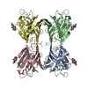
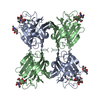
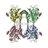
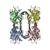
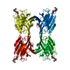
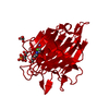
| Title | PHYTOHEMAGGLUTININ-L | ||||||
 Components Components | PHYTOHEMAGGLUTININ-L | ||||||
 Keywords Keywords | LECTIN / GLYCOPROTEIN / PLANT DEFENSE PROTEIN | ||||||
| Function / homology |  Function and homology information Function and homology information | ||||||
| Biological species |  Phaseolus vulgaris (common bean) Phaseolus vulgaris (common bean) | ||||||
| Method |  X-RAY DIFFRACTION / Resolution: 2.8 Å X-RAY DIFFRACTION / Resolution: 2.8 Å | ||||||
 Authors Authors | Hamelryck, T. / Loris, R. | ||||||
 Citation Citation | Journal: J.Biol.Chem. / Year: 1996 Title: The crystallographic structure of phytohemagglutinin-L. Authors: Hamelryck, T.W. / Dao-Thi, M.H. / Poortmans, F. / Chrispeels, M.J. / Wyns, L. / Loris, R. #1: Journal: Proteins / Year: 1996Title: Crystallization of Glycosylated and Nonglycosylated Phytohemagglutinin-L Authors: Dao-Thi, M.H. / Hamelryck, T.W. / Poortmans, F. / Voelker, T.A. / Chrispeels, M.J. / Wyns, L. #2: Journal: Plant Cell / Year: 1991Title: Lectins, Lectin Genes, and Their Role in Plant Defense Authors: Chrispeels, M.J. / Raikhel, N.V. #3: Journal: Embo J. / Year: 1985Title: Characterization of Two Phaseolus Vulgaris Phytohemagglutinin Genes Closely Linked on the Chromosome Authors: Hoffman, L.M. / Donaldson, D.D. | ||||||
| History |
|
- Structure visualization
| Structure viewer | Molecule: MolmilJmol/JSmol |
|---|
- Downloads & links
-Download
| PDBx/mmCIF format | 1fat.cif.gz | 188.8 KB | Display | PDBx/mmCIF format |
|---|---|---|---|---|
| PDB format | pdb1fat.ent.gz | 151.2 KB | Display | PDB format |
| PDBx/mmJSON format | 1fat.json.gz | Tree view | PDBx/mmJSON format | |
| Others |  Other downloads Other downloads |
-Validation report
| Arichive directory | https://data.pdbj.org/pub/pdb/validation_reports/fa/1fatftp://data.pdbj.org/pub/pdb/validation_reports/fa/1fat | HTTPS FTP |
|---|
-Related structure data
| Similar structure data |
|---|


-Links
 PDBj
PDBj

- Assembly
| Deposited unit | 
| ||||||||
|---|---|---|---|---|---|---|---|---|---|
| 1 |
| ||||||||
| Unit cell |
| ||||||||
| Details | PHYTOHEMAGGLUTININ-L IS A HOMOTETRAMER WITH 222 SYMMETRY. EACH SUBUNIT CONSISTS OF 252 RESIDUES. THE FOUR SUBUNITS HAVE CHAIN IDENTIFIERS A, B, C, AND D IN THIS ENTRY. |
-Components
| #1: Protein | Mass: 27438.260 Da / Num. of mol.: 4 / Source method: isolated from a natural source / Details: PURIFIED PHA-L WAS PURCHASED FROM SIGMA / Source: (natural) Phaseolus vulgaris (common bean) / Organ: SEED / References: UniProt: P05087#2: Sugar | ChemComp-NAG /   Type: D-saccharide, beta linking / Mass: 221.208 Da / Num. of mol.: 4 Type: D-saccharide, beta linking / Mass: 221.208 Da / Num. of mol.: 4Source method: isolated from a genetically manipulated source Formula: C8H15NO6 #3: Chemical | ChemComp-MN /   Mass: 54.938 Da / Num. of mol.: 4 / Source method: obtained synthetically / Formula: Mn Mass: 54.938 Da / Num. of mol.: 4 / Source method: obtained synthetically / Formula: Mn#4: Chemical | ChemComp-CA /   Mass: 40.078 Da / Num. of mol.: 4 / Source method: obtained synthetically / Formula: Ca Mass: 40.078 Da / Num. of mol.: 4 / Source method: obtained synthetically / Formula: Ca#5: Water | ChemComp-HOH / |  Mass: 18.015 Da / Num. of mol.: 16 / Source method: isolated from a natural source / Formula: H2O Mass: 18.015 Da / Num. of mol.: 16 / Source method: isolated from a natural source / Formula: H2OHas protein modification | Y | Nonpolymer details | PHYTOHEMAGGLUTININ-L IS A GLYCOPROTEIN. IT CONTAINS ONE HI MANNOSE TYPE AND ONE COMPLEX TYPE SUGAR ...PHYTOHEMAG | |
|---|
-Experimental details
-Experiment
| Experiment | Method: X-RAY DIFFRACTION |
|---|
- Sample preparation
| Crystal | Density Matthews: 2.66 Å3/Da / Density % sol: 45.9 % | ||||||||||||||||||||||||||||||
|---|---|---|---|---|---|---|---|---|---|---|---|---|---|---|---|---|---|---|---|---|---|---|---|---|---|---|---|---|---|---|---|
| Crystal | *PLUS | ||||||||||||||||||||||||||||||
| Crystal grow | *PLUS Temperature: 4 ℃ / pH: 8.5 / Method: vapor diffusion, hanging drop | ||||||||||||||||||||||||||||||
| Components of the solutions | *PLUS
|
-Data collection
| Diffraction source | Wavelength: 1.5418 |
|---|---|
| Detector | Detector: AREA DETECTOR / Date: Jul 5, 1994 |
| Radiation | Monochromatic (M) / Laue (L): M / Scattering type: x-ray |
| Radiation wavelength | Wavelength: 1.5418 Å / Relative weight: 1 |
| Reflection | Redundancy: 2.5 % / Biso Wilson estimate: 43.74 Å2 / Rmerge(I) obs: 0.099 |
- Processing
| Software |
| ||||||||||||||||||||||||||||||||||||||||||||||||||||||||||||
|---|---|---|---|---|---|---|---|---|---|---|---|---|---|---|---|---|---|---|---|---|---|---|---|---|---|---|---|---|---|---|---|---|---|---|---|---|---|---|---|---|---|---|---|---|---|---|---|---|---|---|---|---|---|---|---|---|---|---|---|---|---|
| Refinement | Resolution: 2.8→10 Å / Cross valid method: A POSTERIORI R-FREE FACTOR / σ(F): 0
| ||||||||||||||||||||||||||||||||||||||||||||||||||||||||||||
| Displacement parameters | Biso mean: 24.34 Å2 | ||||||||||||||||||||||||||||||||||||||||||||||||||||||||||||
| Refinement step | Cycle: LAST / Resolution: 2.8→10 Å
| ||||||||||||||||||||||||||||||||||||||||||||||||||||||||||||
| Refine LS restraints |
| ||||||||||||||||||||||||||||||||||||||||||||||||||||||||||||
| Refine LS restraints NCS | NCS model details: MONOMERS A, B, C, D / Weight Biso : 1 / Weight position: 300 | ||||||||||||||||||||||||||||||||||||||||||||||||||||||||||||
| LS refinement shell | Resolution: 2.8→2.94 Å
| ||||||||||||||||||||||||||||||||||||||||||||||||||||||||||||
| Software | *PLUS Name: X-PLOR / Version: 3.1 / Classification: refinement | ||||||||||||||||||||||||||||||||||||||||||||||||||||||||||||
| Refinement | *PLUS Rfactor obs: 0.2 / Rfactor Rwork: 0.2 | ||||||||||||||||||||||||||||||||||||||||||||||||||||||||||||
| Solvent computation | *PLUS | ||||||||||||||||||||||||||||||||||||||||||||||||||||||||||||
| Displacement parameters | *PLUS | ||||||||||||||||||||||||||||||||||||||||||||||||||||||||||||
| Refine LS restraints | *PLUS
|
