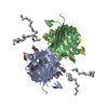
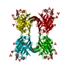
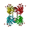
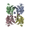
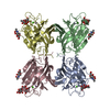
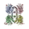
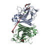
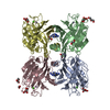
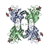
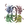
Journal: Int J Biochem Cell Biol / Year: 2016 Title: Structural characterization of a Vatairea macrocarpa lectin in complex with a tumor-associated antigen: A new tool for cancer research. Authors: Bruno L Sousa / José C Silva-Filho / Prashant Kumar / Melissa A Graewert / Ronniery I Pereira / Rodrigo M S Cunha / Kyria S Nascimento / Gustavo A Bezerra / Plínio Delatorre / Kristina ...Authors: Bruno L Sousa / José C Silva-Filho / Prashant Kumar / Melissa A Graewert / Ronniery I Pereira / Rodrigo M S Cunha / Kyria S Nascimento / Gustavo A Bezerra / Plínio Delatorre / Kristina Djinovic-Carugo / Celso S Nagano / Karl Gruber / Benildo S Cavada / Abstract: Legume lectins are the most thoroughly studied group of lectins and have been widely linked to many pathological processes. Their use as immunohistochemistry markers for cell profiling and cancer ...Legume lectins are the most thoroughly studied group of lectins and have been widely linked to many pathological processes. Their use as immunohistochemistry markers for cell profiling and cancer diagnosis have made these molecules important tools for immunological studies and have stimulated the prospection and characterization of new lectins. The crystal structures of a recombinant seed lectin from Vatairea macrocarpa (rVML) and its complexes with GalNAcα1-O-Ser, GalNAc and α-lactose, have been determined at 1.90, 1.97, 2.70 and 1.83Å resolution, respectively. Small angle X-ray scattering and calorimetry assays have confirmed the same pH stable oligomerization pattern and binding profiles proposed for its wild-type counterpart. In silico analyzes have explored the potential of this recombinant lectin as new tool for cancer research through a comparative profile with other legume lectins widely used for cancer diagnosis and prognosis. The results suggest the recognition of specific epitopes exhibited on different cancer cells as a process that relies on the disposition of hydrophobic clusters and charged regions around the lectin carbohydrate-binding site, favouring the anchorage of different groups in the antigen boundaries, highlighting the different potential of each analyzed lectin. In conclusion, the experimental results and comparative analysis show that rVML is as a promising tool for cancer research, able to bind with high affinity specific tumor-associated antigens, highly stable and easily produced.
In the structure databanks used in Yorodumi, some data are registered as the other names, "COVID-19 virus" and "2019-nCoV". Here are the details of the virus and the list of structure data.
Jan 31, 2019. EMDB accession codes are about to change! (news from PDBe EMDB page)
EMDB accession codes are about to change! (news from PDBe EMDB page)
The allocation of 4 digits for EMDB accession codes will soon come to an end. Whilst these codes will remain in use, new EMDB accession codes will include an additional digit and will expand incrementally as the available range of codes is exhausted. The current 4-digit format prefixed with “EMD-” (i.e. EMD-XXXX) will advance to a 5-digit format (i.e. EMD-XXXXX), and so on. It is currently estimated that the 4-digit codes will be depleted around Spring 2019, at which point the 5-digit format will come into force.
The EM Navigator/Yorodumi systems omit the EMD- prefix.
Related info.:Q: What is EMD? / ID/Accession-code notation in Yorodumi/EM Navigator
Yorodumi is a browser for structure data from EMDB, PDB, SASBDB, etc.
This page is also the successor to EM Navigator detail page, and also detail information page/front-end page for Omokage search.
The word "yorodu" (or yorozu) is an old Japanese word meaning "ten thousand". "mi" (miru) is to see.
Related info.:EMDB / PDB / SASBDB / Comparison of 3 databanks / Yorodumi Search / Aug 31, 2016. New EM Navigator & Yorodumi / Yorodumi Papers / Jmol/JSmol / Function and homology information / Changes in new EM Navigator and Yorodumi
 Movie
Movie Controller

 Yorodumi
 Open data
 Basic information
 Components
 Keywords
 Function and homology information
 Vatairea macrocarpa (plant)
 X-RAY DIFFRACTION /
  Authors
 Citation


 Structure visualization
 Downloads & links
 Other downloads
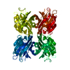

 PDBj
PDBj

 Assembly
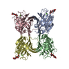


 Mass: 40.078 Da / Num. of mol.: 4 / Source method: obtained synthetically / Formula: Ca
 Mass: 54.938 Da / Num. of mol.: 4 / Source method: obtained synthetically / Formula: Mn
 Mass: 18.015 Da / Num. of mol.: 691 / Source method: isolated from a natural source / Formula: H2O
 Sample preparation
 / Beamline: ID23-2 / Wavelength: 0.872 Å
 Processing