 Movie
Movie Controller

[English] 日本語
 Yorodumi
- PDB-3ul2: Galactose-specific lectin from Dolichos lablab in P6522 space group -
+ Open data

- Basic information
| Entry | Database: PDB / ID: 3ul2 | ||||||
|---|---|---|---|---|---|---|---|
| Title | Galactose-specific lectin from Dolichos lablab in P6522 space group | ||||||
 Components Components | Legume lectin | ||||||
 Keywords Keywords | SUGAR BINDING PROTEIN / Legume lectin fold / Carbohydrate/Sugar-binding / Galactose / Adenine | ||||||
| Function / homology |  Function and homology information Function and homology informationdefense response to fungus / carbohydrate binding / killing of cells of another organism / defense response to bacterium Similarity search - Function | ||||||
| Biological species |  Dolichos lablab (hyacinth bean) Dolichos lablab (hyacinth bean) | ||||||
| Method |  X-RAY DIFFRACTION / SYNCHROTRON / MOLECULAR REPLACEMENT / Resolution: 2.5 Å X-RAY DIFFRACTION / SYNCHROTRON / MOLECULAR REPLACEMENT / Resolution: 2.5 Å | ||||||
 Authors Authors | Shetty, K.N. / Latha, V.L. / Rao, R.N. / Nadimpalli, S.K. / Suguna, K. | ||||||
 Citation Citation | Journal: Iubmb Life / Year: 2013 Title: Affinity of a galactose-specific legume lectin from Dolichos lablab to adenine revealed by X-ray cystallography. Authors: Shetty, K.N. / Latha, V.L. / Rao, R.N. / Nadimpalli, S.K. / Suguna, K. | ||||||
| History |
|
- Structure visualization
| Structure viewer | Molecule: MolmilJmol/JSmol |
|---|
- Downloads & links
-Download
| PDBx/mmCIF format | 3ul2.cif.gz | 204.7 KB | Display | PDBx/mmCIF format |
|---|---|---|---|---|
| PDB format | pdb3ul2.ent.gz | 163.2 KB | Display | PDB format |
| PDBx/mmJSON format | 3ul2.json.gz | Tree view | PDBx/mmJSON format | |
| Others |  Other downloads Other downloads |
-Validation report
| Arichive directory | https://data.pdbj.org/pub/pdb/validation_reports/ul/3ul2ftp://data.pdbj.org/pub/pdb/validation_reports/ul/3ul2 | HTTPS FTP |
|---|
-Related structure data
| Related structure data |  3ujoC  3ujqC  3uk9C  1bjqS S: Starting model for refinement C: citing same article ( |
|---|---|
| Similar structure data |


-Links
 PDBj
PDBj


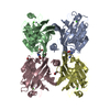
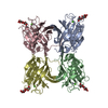
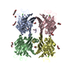
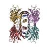
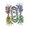
- Assembly
| Deposited unit | 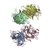
| |||||||||||||||||||||||||||||||||||||||||||||
|---|---|---|---|---|---|---|---|---|---|---|---|---|---|---|---|---|---|---|---|---|---|---|---|---|---|---|---|---|---|---|---|---|---|---|---|---|---|---|---|---|---|---|---|---|---|---|
| 1 | 
| |||||||||||||||||||||||||||||||||||||||||||||
| 2 | 
| |||||||||||||||||||||||||||||||||||||||||||||
| Unit cell |
| |||||||||||||||||||||||||||||||||||||||||||||
| Noncrystallographic symmetry (NCS) | NCS domain:
NCS domain segments:
|
-Components
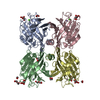
-Protein / Sugars , 2 types, 8 molecules ABCD
| #1: Protein | Mass: 30453.115 Da / Num. of mol.: 4 / Source method: isolated from a natural source / Source: (natural) Dolichos lablab (hyacinth bean) / Tissue: Seed / References: UniProt: B3EWQ9*PLUS#4: Sugar | ChemComp-GAL /  Type: D-saccharide, beta linking / Mass: 180.156 Da / Num. of mol.: 4 Type: D-saccharide, beta linking / Mass: 180.156 Da / Num. of mol.: 4Source method: isolated from a genetically manipulated source Formula: C6H12O6 |
|---|
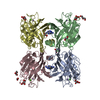
-Non-polymers , 5 types, 185 molecules 


| #2: Chemical | ChemComp-CA /  Mass: 40.078 Da / Num. of mol.: 4 / Source method: obtained synthetically / Formula: Ca Mass: 40.078 Da / Num. of mol.: 4 / Source method: obtained synthetically / Formula: Ca#3: Chemical | ChemComp-MN /  Mass: 54.938 Da / Num. of mol.: 4 / Source method: obtained synthetically / Formula: Mn Mass: 54.938 Da / Num. of mol.: 4 / Source method: obtained synthetically / Formula: Mn#5: Chemical | ChemComp-EDO /  Mass: 62.068 Da / Num. of mol.: 8 / Source method: obtained synthetically / Formula: C2H6O2 Mass: 62.068 Da / Num. of mol.: 8 / Source method: obtained synthetically / Formula: C2H6O2#6: Chemical |  Mass: 106.120 Da / Num. of mol.: 2 / Source method: obtained synthetically / Formula: C4H10O3 Mass: 106.120 Da / Num. of mol.: 2 / Source method: obtained synthetically / Formula: C4H10O3#7: Water | ChemComp-HOH / | Mass: 18.015 Da / Num. of mol.: 167 / Source method: isolated from a natural source / Formula: H2O |
|---|
-Details
| Sequence details | A SEQUENCE DATABASE REFERENCE FOR THIS PROTEIN DOES NOT CURRENTLY EXIST. |
|---|
-Experimental details
-Experiment
| Experiment | Method: X-RAY DIFFRACTION / Number of used crystals: 1 |
|---|
- Sample preparation
| Crystal | Density Matthews: 2.5 Å3/Da / Density % sol: 55.75 % |
|---|---|
| Crystal grow | Temperature: 289 K / Method: vapor diffusion, sitting drop / pH: 7.5 Details: 30% PEG 4K, 100mM Tris, 200mM Lithium sulphate , pH 7.5, VAPOR DIFFUSION, SITTING DROP, temperature 289K |
-Data collection
| Diffraction | Mean temperature: 100 K |
|---|---|
| Diffraction source | Source: SYNCHROTRON / Site: ESRF  / Beamline: BM14 / Wavelength: 0.97 Å / Beamline: BM14 / Wavelength: 0.97 Å |
| Detector | Type: MARMOSAIC 300 mm CCD / Detector: CCD / Date: Jul 24, 2010 |
| Radiation | Protocol: SINGLE WAVELENGTH / Monochromatic (M) / Laue (L): M / Scattering type: x-ray |
| Radiation wavelength | Wavelength: 0.97 Å / Relative weight: 1 |
| Reflection | Resolution: 2.5→50 Å / Num. obs: 44836 / % possible obs: 93.4 % / Redundancy: 9.7 % / Biso Wilson estimate: 57.7 Å2 / Rmerge(I) obs: 0.101 / Net I/σ(I): 27.1 |
| Reflection shell | Resolution: 2.5→2.54 Å / Redundancy: 4.8 % / Rmerge(I) obs: 0.448 / Mean I/σ(I) obs: 2.7 / Num. unique all: 2032 / % possible all: 95.7 |
- Processing
| Software |
| |||||||||||||||||||||||||||||||||||||||||||||||||||||||||||||||||||||||||||||||||||||
|---|---|---|---|---|---|---|---|---|---|---|---|---|---|---|---|---|---|---|---|---|---|---|---|---|---|---|---|---|---|---|---|---|---|---|---|---|---|---|---|---|---|---|---|---|---|---|---|---|---|---|---|---|---|---|---|---|---|---|---|---|---|---|---|---|---|---|---|---|---|---|---|---|---|---|---|---|---|---|---|---|---|---|---|---|---|---|
| Refinement | Method to determine structure: MOLECULAR REPLACEMENT Starting model: 1BJQ Resolution: 2.5→50 Å / Cor.coef. Fo:Fc: 0.886 / Cor.coef. Fo:Fc free: 0.85 / SU B: 11.158 / SU ML: 0.253 / Cross valid method: THROUGHOUT / ESU R Free: 0.337 / Stereochemistry target values: MAXIMUM LIKELIHOOD / Details: HYDROGENS HAVE BEEN ADDED IN THE RIDING POSITIONS
| |||||||||||||||||||||||||||||||||||||||||||||||||||||||||||||||||||||||||||||||||||||
| Solvent computation | Ion probe radii: 0.8 Å / Shrinkage radii: 0.8 Å / VDW probe radii: 1.4 Å / Solvent model: MASK | |||||||||||||||||||||||||||||||||||||||||||||||||||||||||||||||||||||||||||||||||||||
| Displacement parameters | Biso mean: 37.259 Å2
| |||||||||||||||||||||||||||||||||||||||||||||||||||||||||||||||||||||||||||||||||||||
| Refinement step | Cycle: LAST / Resolution: 2.5→50 Å
| |||||||||||||||||||||||||||||||||||||||||||||||||||||||||||||||||||||||||||||||||||||
| Refine LS restraints |
| |||||||||||||||||||||||||||||||||||||||||||||||||||||||||||||||||||||||||||||||||||||
| Refine LS restraints NCS | Dom-ID: 1 / Ens-ID: 1 / Refine-ID: X-RAY DIFFRACTION
| |||||||||||||||||||||||||||||||||||||||||||||||||||||||||||||||||||||||||||||||||||||
| LS refinement shell | Resolution: 2.502→2.567 Å / Total num. of bins used: 20
|
