 Movie
Movie Controller

[English] 日本語
 Yorodumi
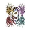
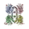
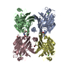
- PDB-1lu2: DOLICHOS BIFLORUS SEED LECTIN IN COMPLEX WITH THE BLOOD GROUP A T... -
+ Open data

- Basic information
| Entry | Database: PDB / ID: 1lu2 | |||||||||
|---|---|---|---|---|---|---|---|---|---|---|
| Title | DOLICHOS BIFLORUS SEED LECTIN IN COMPLEX WITH THE BLOOD GROUP A TRISACCHARIDE | |||||||||
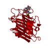
 Components Components | LECTIN | |||||||||
 Keywords Keywords | LECTIN / LEGUME LECTINS / DOLICHOS BIFLORUS SEED LECTIN / SUGAR BINDING | |||||||||
| Function / homology |  Function and homology information Function and homology information | |||||||||
| Biological species |  Vigna unguiculata subsp. cylindrica (catjang cowpea) Vigna unguiculata subsp. cylindrica (catjang cowpea) | |||||||||
| Method |  X-RAY DIFFRACTION / MOLECULAR REPLACEMENT / Resolution: 2.8 Å X-RAY DIFFRACTION / MOLECULAR REPLACEMENT / Resolution: 2.8 Å | |||||||||
 Authors Authors | Hamelryck, T.W. / Loris, R. / Bouckaert, J. / Strecker, G. / Imberty, A. / Fernandez, E. / Wyns, L. / Etzler, M.E. | |||||||||
 Citation Citation | Journal: J.Mol.Biol. / Year: 1999 Title: Carbohydrate binding, quaternary structure and a novel hydrophobic binding site in two legume lectin oligomers from Dolichos biflorus. Authors: Hamelryck, T.W. / Loris, R. / Bouckaert, J. / Dao-Thi, M.H. / Strecker, G. / Imberty, A. / Fernandez, E. / Wyns, L. / Etzler, M.E. | |||||||||
| History |
|
- Structure visualization
| Structure viewer | Molecule: MolmilJmol/JSmol |
|---|
- Downloads & links
-Download
| PDBx/mmCIF format | 1lu2.cif.gz | 105.3 KB | Display | PDBx/mmCIF format |
|---|---|---|---|---|
| PDB format | pdb1lu2.ent.gz | 80.9 KB | Display | PDB format |
| PDBx/mmJSON format | 1lu2.json.gz | Tree view | PDBx/mmJSON format | |
| Others |  Other downloads Other downloads |
-Validation report
| Arichive directory | https://data.pdbj.org/pub/pdb/validation_reports/lu/1lu2ftp://data.pdbj.org/pub/pdb/validation_reports/lu/1lu2 | HTTPS FTP |
|---|
-Related structure data
| Related structure data | 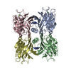 1bjqC  1lu1C 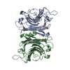 1lulC  1fatS S: Starting model for refinement C: citing same article ( |
|---|---|
| Similar structure data |


-Links
 PDBj
PDBj

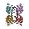
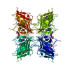
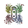
- Assembly
| Deposited unit | 
| ||||||||
|---|---|---|---|---|---|---|---|---|---|
| 1 | 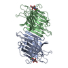
| ||||||||
| Unit cell |
| ||||||||
| Noncrystallographic symmetry (NCS) | NCS oper: (Code: given Matrix: (-0.9999, 0.0016, 0.0136), Vector: |
-Components
| #1: Protein | Mass: 27115.078 Da / Num. of mol.: 2 Source method: isolated from a genetically manipulated source Details: DOLICHOS BIFLORUS SEED LECTIN Source: (gene. exp.) Vigna unguiculata subsp. cylindrica (catjang cowpea)Species: Vigna unguiculata / Strain: subsp. cylindrica / Organ: SEED / Production host:  #2: Sugar |   Type: D-saccharide, alpha linking / Mass: 221.208 Da / Num. of mol.: 2 Type: D-saccharide, alpha linking / Mass: 221.208 Da / Num. of mol.: 2Source method: isolated from a genetically manipulated source Formula: C8H15NO6 #3: Chemical |   Mass: 40.078 Da / Num. of mol.: 2 / Source method: obtained synthetically / Formula: Ca Mass: 40.078 Da / Num. of mol.: 2 / Source method: obtained synthetically / Formula: Ca#4: Chemical |   Mass: 54.938 Da / Num. of mol.: 2 / Source method: obtained synthetically / Formula: Mn Mass: 54.938 Da / Num. of mol.: 2 / Source method: obtained synthetically / Formula: Mn |
|---|
-Experimental details
-Experiment
| Experiment | Method: X-RAY DIFFRACTION / Number of used crystals: 1 |
|---|
- Sample preparation
| Crystal | Density Matthews: 2.6 Å3/Da / Density % sol: 55 % | |||||||||||||||||||||||||
|---|---|---|---|---|---|---|---|---|---|---|---|---|---|---|---|---|---|---|---|---|---|---|---|---|---|---|
| Crystal grow | pH: 6.6 Details: 100 MM NACACO PH 6.6 15% (W/V) PEG-ME 5000 10 MM BLOOD GROUP A TRISACCHARIDE | |||||||||||||||||||||||||
| Crystal grow | *PLUS Method: vapor diffusion | |||||||||||||||||||||||||
| Components of the solutions | *PLUS
|
-Data collection
| Diffraction | Mean temperature: 293 K |
|---|---|
| Diffraction source | Source: ROTATING ANODE / Type: RIGAKU / Wavelength: 1.5418 |
| Detector | Type: MARRESEARCH / Detector: IMAGE PLATE / Date: Jan 1, 1998 |
| Radiation | Monochromatic (M) / Laue (L): M / Scattering type: x-ray |
| Radiation wavelength | Wavelength: 1.5418 Å / Relative weight: 1 |
| Reflection | Resolution: 2.8→15 Å / Num. obs: 14331 / % possible obs: 85.2 % / Observed criterion σ(I): 0 / Redundancy: 2.7 % / Biso Wilson estimate: 29.7 Å2 / Rmerge(I) obs: 0.11 / Net I/σ(I): 10.7 |
| Reflection shell | Resolution: 2.8→2.91 Å / Redundancy: 1.8 % / Rmerge(I) obs: 0.274 / Mean I/σ(I) obs: 3.1 / % possible all: 85.8 |
| Reflection | *PLUS Num. measured all: 39403 |
| Reflection shell | *PLUS Lowest resolution: 2.92 Å |
- Processing
| Software |
| ||||||||||||||||||||||||||||||||||||||||||||||||||||||||||||||||||||||||||||||||
|---|---|---|---|---|---|---|---|---|---|---|---|---|---|---|---|---|---|---|---|---|---|---|---|---|---|---|---|---|---|---|---|---|---|---|---|---|---|---|---|---|---|---|---|---|---|---|---|---|---|---|---|---|---|---|---|---|---|---|---|---|---|---|---|---|---|---|---|---|---|---|---|---|---|---|---|---|---|---|---|---|---|
| Refinement | Method to determine structure: MOLECULAR REPLACEMENT Starting model: PDB ENTRY 1FAT Resolution: 2.8→15 Å / Rfactor Rfree error: 0.006 / Data cutoff high absF: 10000000 / Data cutoff low absF: 0.001 / Isotropic thermal model: RESTRAINED / Cross valid method: THROUGHOUT / σ(F): 0 Details: BULK SOLVENT MODEL USED ONLY THE GALNAC RESIDUE OF THE BLOOD GROUP A TRISACCHARIDE IS VISIBLE IN THE ELECTRON DENSITY.
| ||||||||||||||||||||||||||||||||||||||||||||||||||||||||||||||||||||||||||||||||
| Displacement parameters | Biso mean: 32.1 Å2
| ||||||||||||||||||||||||||||||||||||||||||||||||||||||||||||||||||||||||||||||||
| Refine analyze |
| ||||||||||||||||||||||||||||||||||||||||||||||||||||||||||||||||||||||||||||||||
| Refinement step | Cycle: LAST / Resolution: 2.8→15 Å
| ||||||||||||||||||||||||||||||||||||||||||||||||||||||||||||||||||||||||||||||||
| Refine LS restraints |
| ||||||||||||||||||||||||||||||||||||||||||||||||||||||||||||||||||||||||||||||||
| LS refinement shell | Resolution: 2.8→2.9 Å / Rfactor Rfree error: 0.027 / Total num. of bins used: 10
| ||||||||||||||||||||||||||||||||||||||||||||||||||||||||||||||||||||||||||||||||
| Xplor file |
| ||||||||||||||||||||||||||||||||||||||||||||||||||||||||||||||||||||||||||||||||
| Software | *PLUS Name: X-PLOR / Version: 3.851 / Classification: refinement | ||||||||||||||||||||||||||||||||||||||||||||||||||||||||||||||||||||||||||||||||
| Refinement | *PLUS | ||||||||||||||||||||||||||||||||||||||||||||||||||||||||||||||||||||||||||||||||
| Solvent computation | *PLUS | ||||||||||||||||||||||||||||||||||||||||||||||||||||||||||||||||||||||||||||||||
| Displacement parameters | *PLUS | ||||||||||||||||||||||||||||||||||||||||||||||||||||||||||||||||||||||||||||||||
| Refine LS restraints | *PLUS
| ||||||||||||||||||||||||||||||||||||||||||||||||||||||||||||||||||||||||||||||||
| LS refinement shell | *PLUS Rfactor obs: 0.289 |
