 Movie
Movie Controller

[English] 日本語
 Yorodumi
- PDB-3ega: Crystal structure of Pellino2 FHA Domain at 1.8 Angstroms resolution -
+ Open data

- Basic information
| Entry | Database: PDB / ID: 3ega | ||||||
|---|---|---|---|---|---|---|---|
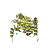
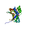
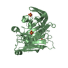
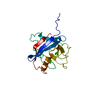
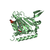
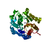
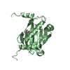
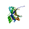
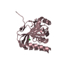
| Title | Crystal structure of Pellino2 FHA Domain at 1.8 Angstroms resolution | ||||||
 Components Components | Protein pellino homolog 2 | ||||||
 Keywords Keywords | PROTEIN BINDING / Pellino / FHA Domain / E3 ubiquitin ligase / substrate binding domain / Phosphoprotein | ||||||
| Function / homology |  Function and homology information Function and homology informationregulation of Toll signaling pathway / ubiquitin-ubiquitin ligase activity / IRAK1 recruits IKK complex / IRAK1 recruits IKK complex upon TLR7/8 or 9 stimulation / positive regulation of protein phosphorylation / RING-type E3 ubiquitin transferase / Interleukin-1 signaling / protein polyubiquitination / ubiquitin protein ligase activity / positive regulation of canonical NF-kappaB signal transduction ...regulation of Toll signaling pathway / ubiquitin-ubiquitin ligase activity / IRAK1 recruits IKK complex / IRAK1 recruits IKK complex upon TLR7/8 or 9 stimulation / positive regulation of protein phosphorylation / RING-type E3 ubiquitin transferase / Interleukin-1 signaling / protein polyubiquitination / ubiquitin protein ligase activity / positive regulation of canonical NF-kappaB signal transduction / positive regulation of MAPK cascade / cytosol Similarity search - Function | ||||||
| Biological species |  Homo sapiens (human) Homo sapiens (human) | ||||||
| Method |  X-RAY DIFFRACTION / SYNCHROTRON / MAD / Resolution: 1.8 Å X-RAY DIFFRACTION / SYNCHROTRON / MAD / Resolution: 1.8 Å | ||||||
 Authors Authors | Ferguson, K.M. / Lin, C. / Schmitz, K.R. | ||||||
 Citation Citation | Journal: Structure / Year: 2008 Title: Pellino proteins contain a cryptic FHA domain that mediates interaction with phosphorylated IRAK1. Authors: Lin, C.C. / Huoh, Y.S. / Schmitz, K.R. / Jensen, L.E. / Ferguson, K.M. | ||||||
| History |
|
- Structure visualization
| Structure viewer | Molecule: MolmilJmol/JSmol |
|---|
- Downloads & links
-Download
| PDBx/mmCIF format | 3ega.cif.gz | 61.3 KB | Display | PDBx/mmCIF format |
|---|---|---|---|---|
| PDB format | pdb3ega.ent.gz | 43.4 KB | Display | PDB format |
| PDBx/mmJSON format | 3ega.json.gz | Tree view | PDBx/mmJSON format | |
| Others |  Other downloads Other downloads |
-Validation report
| Arichive directory | https://data.pdbj.org/pub/pdb/validation_reports/eg/3egaftp://data.pdbj.org/pub/pdb/validation_reports/eg/3ega | HTTPS FTP |
|---|
-Related structure data


-Links
 PDBj
PDBj

- Assembly
| Deposited unit | 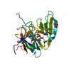
| ||||||||
|---|---|---|---|---|---|---|---|---|---|
| 1 |
| ||||||||
| 2 | 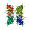
| ||||||||
| Unit cell |
| ||||||||
| Components on special symmetry positions |
|
-Components
| #1: Protein | Mass: 29319.123 Da / Num. of mol.: 1 / Fragment: UNP residues 15-275, FHA domain / Mutation: V61M, L232M Source method: isolated from a genetically manipulated source Source: (gene. exp.) Homo sapiens (human) / Gene: PELI2 / Plasmid: pET28 derivative (HTUA) / Production host:  |
|---|---|
| #2: Chemical | ChemComp-SO4 /   Mass: 96.063 Da / Num. of mol.: 1 / Source method: obtained synthetically / Formula: SO4 Mass: 96.063 Da / Num. of mol.: 1 / Source method: obtained synthetically / Formula: SO4 |
| #3: Water | ChemComp-HOH /  Mass: 18.015 Da / Num. of mol.: 128 / Source method: isolated from a natural source / Formula: H2O Mass: 18.015 Da / Num. of mol.: 128 / Source method: isolated from a natural source / Formula: H2O |
| Has protein modification | Y |
-Experimental details
-Experiment
| Experiment | Method: X-RAY DIFFRACTION / Number of used crystals: 1 |
|---|
- Sample preparation
| Crystal | Density Matthews: 2.5 Å3/Da / Density % sol: 50.8 % |
|---|---|
| Crystal grow | Temperature: 295 K / Method: vapor diffusion / pH: 5.5 Details: 0.1 M sodium acetate, 26.4% w/v PEG 2000 MME, 0.2 M ammonium sulfate, pH 5.5, vapor diffusion, temperature 295K |
-Data collection
| Diffraction | Mean temperature: 100 K | |||||||||||||||||||||||||||||||||||||||||||||||||||||||||||||||||||||||||||||
|---|---|---|---|---|---|---|---|---|---|---|---|---|---|---|---|---|---|---|---|---|---|---|---|---|---|---|---|---|---|---|---|---|---|---|---|---|---|---|---|---|---|---|---|---|---|---|---|---|---|---|---|---|---|---|---|---|---|---|---|---|---|---|---|---|---|---|---|---|---|---|---|---|---|---|---|---|---|---|
| Diffraction source | Source: SYNCHROTRON / Site: APS  / Beamline: 23-ID-B / Wavelength: 0.9795, 0.9796, 0.9495 / Beamline: 23-ID-B / Wavelength: 0.9795, 0.9796, 0.9495 | |||||||||||||||||||||||||||||||||||||||||||||||||||||||||||||||||||||||||||||
| Detector | Type: ADSC QUANTUM 210 / Detector: CCD / Date: Aug 5, 2007 | |||||||||||||||||||||||||||||||||||||||||||||||||||||||||||||||||||||||||||||
| Radiation | Protocol: MAD / Monochromatic (M) / Laue (L): M / Scattering type: x-ray | |||||||||||||||||||||||||||||||||||||||||||||||||||||||||||||||||||||||||||||
| Radiation wavelength |
| |||||||||||||||||||||||||||||||||||||||||||||||||||||||||||||||||||||||||||||
| Reflection | Redundancy: 8.2 % / Av σ(I) over netI: 37.26 / Number: 221150 / Rmerge(I) obs: 0.1 / Χ2: 3.47 / D res high: 1.8 Å / D res low: 50 Å / Num. obs: 27082 / % possible obs: 98.5 | |||||||||||||||||||||||||||||||||||||||||||||||||||||||||||||||||||||||||||||
| Diffraction reflection shell |
| |||||||||||||||||||||||||||||||||||||||||||||||||||||||||||||||||||||||||||||
| Reflection | Resolution: 1.8→50 Å / Num. all: 27082 / Num. obs: 27082 / % possible obs: 98.5 % / Observed criterion σ(F): 0 / Observed criterion σ(I): 0 / Redundancy: 8.2 % / Rmerge(I) obs: 0.1 / Χ2: 3.469 / Net I/σ(I): 37.257 | |||||||||||||||||||||||||||||||||||||||||||||||||||||||||||||||||||||||||||||
| Reflection shell | Resolution: 1.8→1.86 Å / Redundancy: 3.9 % / Rmerge(I) obs: 0.515 / Mean I/σ(I) obs: 4 / Num. unique all: 2345 / Χ2: 0.881 / % possible all: 86.9 |
-Phasing
| Phasing | Method: MAD |
|---|
- Processing
| Software |
| ||||||||||||||||||||||||||||||||||||||||||||||||||||||||||||||||||||||||||||||||||||||||||
|---|---|---|---|---|---|---|---|---|---|---|---|---|---|---|---|---|---|---|---|---|---|---|---|---|---|---|---|---|---|---|---|---|---|---|---|---|---|---|---|---|---|---|---|---|---|---|---|---|---|---|---|---|---|---|---|---|---|---|---|---|---|---|---|---|---|---|---|---|---|---|---|---|---|---|---|---|---|---|---|---|---|---|---|---|---|---|---|---|---|---|---|
| Refinement | Method to determine structure: MAD / Resolution: 1.8→43.07 Å / Cor.coef. Fo:Fc: 0.946 / Cor.coef. Fo:Fc free: 0.937 / WRfactor Rfree: 0.243 / WRfactor Rwork: 0.211 / Occupancy max: 1 / Occupancy min: 0.5 / FOM work R set: 0.968 / SU B: 2.335 / SU ML: 0.075 / SU R Cruickshank DPI: 0.122 / SU Rfree: 0.12 / Cross valid method: THROUGHOUT / σ(F): 0 / σ(I): 0 / ESU R: 0.122 / ESU R Free: 0.12 Stereochemistry target values: MAXIMUM LIKELIHOOD WITH PHASES Details: HYDROGENS HAVE BEEN ADDED IN THE RIDING POSITIONS
| ||||||||||||||||||||||||||||||||||||||||||||||||||||||||||||||||||||||||||||||||||||||||||
| Solvent computation | Ion probe radii: 0.8 Å / Shrinkage radii: 0.8 Å / VDW probe radii: 1.2 Å / Solvent model: MASK | ||||||||||||||||||||||||||||||||||||||||||||||||||||||||||||||||||||||||||||||||||||||||||
| Displacement parameters | Biso max: 54.52 Å2 / Biso mean: 25.985 Å2 / Biso min: 6.96 Å2
| ||||||||||||||||||||||||||||||||||||||||||||||||||||||||||||||||||||||||||||||||||||||||||
| Refinement step | Cycle: LAST / Resolution: 1.8→43.07 Å
| ||||||||||||||||||||||||||||||||||||||||||||||||||||||||||||||||||||||||||||||||||||||||||
| Refine LS restraints |
| ||||||||||||||||||||||||||||||||||||||||||||||||||||||||||||||||||||||||||||||||||||||||||
| LS refinement shell | Resolution: 1.8→1.843 Å / Total num. of bins used: 20
|
