 Movie
Movie Controller


+ Open data

- Basic information
| Entry | Database: PDB / ID: 1lul | ||||||
|---|---|---|---|---|---|---|---|
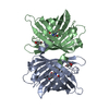
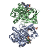
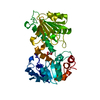
| Title | DB58, A LEGUME LECTIN FROM DOLICHOS BIFLORUS | ||||||
 Components Components | LECTIN DB58 | ||||||
 Keywords Keywords | LECTIN / LEGUME LECTIN / QUATERNARY STRUCTURE / PLANT HORMONE BINDING / GLYCOPROTEIN | ||||||
| Function / homology |  Function and homology information Function and homology information | ||||||
| Biological species |  Vigna unguiculata subsp. cylindrica (catjang cowpea) Vigna unguiculata subsp. cylindrica (catjang cowpea) | ||||||
| Method |  X-RAY DIFFRACTION / SYNCHROTRON / MOLECULAR REPLACEMENT / Resolution: 3.3 Å X-RAY DIFFRACTION / SYNCHROTRON / MOLECULAR REPLACEMENT / Resolution: 3.3 Å | ||||||
 Authors Authors | Hamelryck, T.W. / Bouckaert, J. / Dao-Thi, M.H. / Wyns, L. / Etzler, M. / Loris, R. | ||||||
 Citation Citation | Journal: J.Mol.Biol. / Year: 1999 Title: Carbohydrate binding, quaternary structure and a novel hydrophobic binding site in two legume lectin oligomers from Dolichos biflorus. Authors: Hamelryck, T.W. / Loris, R. / Bouckaert, J. / Dao-Thi, M.H. / Strecker, G. / Imberty, A. / Fernandez, E. / Wyns, L. / Etzler, M.E. #1: Journal: Acta Crystallogr.,Sect.D / Year: 1998Title: Crystallization of Two Related Lectins from the Legume Plant Dolichos Biflorus Authors: Dao-Thi, M.-H. / Hamelryck, T.W. / Bouckaert, J. / Korber, F. / Burkow, V. / Poortmans, F. / Etzler, M. / Strecker, G. / Wyns, L. / Loris, R. | ||||||
| History |
|
- Structure visualization
| Structure viewer | Molecule: MolmilJmol/JSmol |
|---|
- Downloads & links
-Download
| PDBx/mmCIF format | 1lul.cif.gz | 268.2 KB | Display | PDBx/mmCIF format |
|---|---|---|---|---|
| PDB format | pdb1lul.ent.gz | 216.4 KB | Display | PDB format |
| PDBx/mmJSON format | 1lul.json.gz | Tree view | PDBx/mmJSON format | |
| Others |  Other downloads Other downloads |
-Validation report
| Arichive directory | https://data.pdbj.org/pub/pdb/validation_reports/lu/1lulftp://data.pdbj.org/pub/pdb/validation_reports/lu/1lul | HTTPS FTP |
|---|
-Related structure data
| Related structure data |  1bjqSC 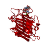 1lu1C 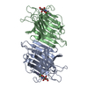 1lu2C S: Starting model for refinement C: citing same article ( |
|---|---|
| Similar structure data |


-Links
 PDBj
PDBj

- Assembly
| Deposited unit | 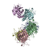
| ||||||||||||||||||||||||
|---|---|---|---|---|---|---|---|---|---|---|---|---|---|---|---|---|---|---|---|---|---|---|---|---|---|
| 1 | 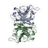
| ||||||||||||||||||||||||
| 2 | 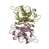
| ||||||||||||||||||||||||
| 3 | 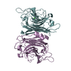
| ||||||||||||||||||||||||
| Unit cell |
| ||||||||||||||||||||||||
| Noncrystallographic symmetry (NCS) | NCS oper:
| ||||||||||||||||||||||||
| Details | DB58 IS A LEGUME LECTIN FROM THE STEMS AND THE LEAVES OF THE TROPICAL LEGUME PLANT DOLICHOS BIFLORUS. IT IS A HETERODIMER THAT CONSISTS OF A TRUNCATED (241 AA) AND AN INTACT SUBUNIT (253 AA). THIS A LOW RESOLUTION STRUCTURE (3.3 ANGSTROM) AND SHOULD BE VIEWED WITH CAUTION. HOWEVER, THE STRUCTURE SHOWS CLEARLY THAT DB58 REPRESENTS THE FOURTH KNOWN LEGUME LECTIN DIMER TYPE, RELATED TO THE PHA-L TYPE TETRAMER. LAST SIX RESIDUES ARE NOT VISIBLE IN THE DENSITY. |
-Components
| #1: Protein | Mass: 27146.885 Da / Num. of mol.: 6 / Source method: isolated from a natural source Source: (natural) Vigna unguiculata subsp. cylindrica (catjang cowpea)Organ: STEM AND LEAF / Species: Vigna unguiculata / Strain: subsp. cylindrica / References: UniProt: P19588 #2: Chemical | ChemComp-MN /   Mass: 54.938 Da / Num. of mol.: 6 / Source method: obtained synthetically / Formula: Mn Mass: 54.938 Da / Num. of mol.: 6 / Source method: obtained synthetically / Formula: Mn#3: Chemical | ChemComp-CA /   Mass: 40.078 Da / Num. of mol.: 6 / Source method: obtained synthetically / Formula: Ca Mass: 40.078 Da / Num. of mol.: 6 / Source method: obtained synthetically / Formula: Ca |
|---|
-Experimental details
-Experiment
| Experiment | Method: X-RAY DIFFRACTION / Number of used crystals: 1 |
|---|
- Sample preparation
| Crystal | Density Matthews: 2.6 Å3/Da / Density % sol: 48.1 % | ||||||||||||||||||||||||||||||
|---|---|---|---|---|---|---|---|---|---|---|---|---|---|---|---|---|---|---|---|---|---|---|---|---|---|---|---|---|---|---|---|
| Crystal grow | pH: 5.6 Details: 100 MM NA-CITRATE PH 5.6 10 % (W/V) PEG 6000 0.3 M NACL | ||||||||||||||||||||||||||||||
| Crystal grow | *PLUS Method: vapor diffusion, hanging drop | ||||||||||||||||||||||||||||||
| Components of the solutions | *PLUS
|
-Data collection
| Diffraction | Mean temperature: 293 K |
|---|---|
| Diffraction source | Source: SYNCHROTRON / Site: SRS  / Beamline: PX7.2 / Wavelength: 1.488 / Beamline: PX7.2 / Wavelength: 1.488 |
| Detector | Type: MARRESEARCH / Detector: IMAGE PLATE / Date: Mar 1, 1998 / Details: A/A |
| Radiation | Monochromatic (M) / Laue (L): M / Scattering type: x-ray |
| Radiation wavelength | Wavelength: 1.488 Å / Relative weight: 1 |
| Reflection | Resolution: 3.3→20 Å / Num. obs: 22745 / % possible obs: 80.6 % / Observed criterion σ(I): 0 / Redundancy: 2.07 % / Biso Wilson estimate: 28.6 Å2 / Rmerge(I) obs: 0.135 / Net I/σ(I): 10.2 |
| Reflection shell | Resolution: 3.3→3.42 Å / Redundancy: 1.7 % / Rmerge(I) obs: 0.279 / Mean I/σ(I) obs: 5.5 / % possible all: 68 |
| Reflection | *PLUS Num. measured all: 47794 |
| Reflection shell | *PLUS Lowest resolution: 3.33 Å / Mean I/σ(I) obs: 2.7 |
- Processing
| Software |
| ||||||||||||||||||||||||||||||||||||||||||||||||||||||||||||||||||||||||||||||||
|---|---|---|---|---|---|---|---|---|---|---|---|---|---|---|---|---|---|---|---|---|---|---|---|---|---|---|---|---|---|---|---|---|---|---|---|---|---|---|---|---|---|---|---|---|---|---|---|---|---|---|---|---|---|---|---|---|---|---|---|---|---|---|---|---|---|---|---|---|---|---|---|---|---|---|---|---|---|---|---|---|---|
| Refinement | Method to determine structure: MOLECULAR REPLACEMENT Starting model: PDB ENTRY 1BJQ Resolution: 3.3→20 Å / Rfactor Rfree error: 0.006 / Data cutoff high absF: 10000000 / Data cutoff low absF: 0.001 / Isotropic thermal model: CONSTR / Cross valid method: THROUGHOUT / σ(F): 0
| ||||||||||||||||||||||||||||||||||||||||||||||||||||||||||||||||||||||||||||||||
| Displacement parameters | Biso mean: 53.6 Å2
| ||||||||||||||||||||||||||||||||||||||||||||||||||||||||||||||||||||||||||||||||
| Refine analyze |
| ||||||||||||||||||||||||||||||||||||||||||||||||||||||||||||||||||||||||||||||||
| Refinement step | Cycle: LAST / Resolution: 3.3→20 Å
| ||||||||||||||||||||||||||||||||||||||||||||||||||||||||||||||||||||||||||||||||
| Refine LS restraints |
| ||||||||||||||||||||||||||||||||||||||||||||||||||||||||||||||||||||||||||||||||
| Refine LS restraints NCS | NCS model details: RESTRAINTS | ||||||||||||||||||||||||||||||||||||||||||||||||||||||||||||||||||||||||||||||||
| LS refinement shell | Resolution: 3.3→3.42 Å / Rfactor Rfree error: 0.03 / Total num. of bins used: 10
| ||||||||||||||||||||||||||||||||||||||||||||||||||||||||||||||||||||||||||||||||
| Xplor file |
| ||||||||||||||||||||||||||||||||||||||||||||||||||||||||||||||||||||||||||||||||
| Software | *PLUS Name: X-PLOR / Version: 3.851 / Classification: refinement | ||||||||||||||||||||||||||||||||||||||||||||||||||||||||||||||||||||||||||||||||
| Refinement | *PLUS Rfactor obs: 0.227 | ||||||||||||||||||||||||||||||||||||||||||||||||||||||||||||||||||||||||||||||||
| Solvent computation | *PLUS | ||||||||||||||||||||||||||||||||||||||||||||||||||||||||||||||||||||||||||||||||
| Displacement parameters | *PLUS | ||||||||||||||||||||||||||||||||||||||||||||||||||||||||||||||||||||||||||||||||
| Refine LS restraints | *PLUS
| ||||||||||||||||||||||||||||||||||||||||||||||||||||||||||||||||||||||||||||||||
| LS refinement shell | *PLUS Rfactor obs: 0.304 |
