 Movie
Movie Controller

[English] 日本語
 Yorodumi
- PDB-1fnz: A bark lectin from robinia pseudoacacia in complex with N-acetylg... -
+ Open data

- Basic information
| Entry | Database: PDB / ID: 1fnz | |||||||||
|---|---|---|---|---|---|---|---|---|---|---|
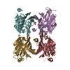
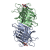
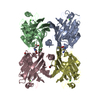
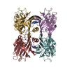
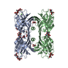
| Title | A bark lectin from robinia pseudoacacia in complex with N-acetylgalactosamine | |||||||||
 Components Components | BARK AGGLUTININ I, POLYPEPTIDE A | |||||||||
 Keywords Keywords | SUGAR BINDING PROTEIN / jelly roll | |||||||||
| Function / homology |  Function and homology information Function and homology information | |||||||||
| Biological species |  Robinia pseudoacacia (black locust) Robinia pseudoacacia (black locust) | |||||||||
| Method |  X-RAY DIFFRACTION / SYNCHROTRON / Resolution: 2.05 Å X-RAY DIFFRACTION / SYNCHROTRON / Resolution: 2.05 Å | |||||||||
 Authors Authors | Rabijns, A. / Verboven, C. / Rouge, P. / Barre, A. / Van Damme, E.J. / Peumans, W.J. / De Ranter, C.J. | |||||||||
 Citation Citation | Journal: Proteins / Year: 2001 Title: Structure of a legume lectin from the bark of Robinia pseudoacacia and its complex with N-acetylgalactosamine Authors: Rabijns, A. / Verboven, C. / Rouge, P. / Barre, A. / Van Damme, E.J. / Peumans, W.J. / De Ranter, C.J. #1: Journal: Acta Crystallogr.,Sect.D / Year: 2000Title: A legume lectin from the bark of Robinia pseudoacacia crystallizes in two crystal forms: preliminary diffraction analyses Authors: Rabijns, A. / Verboven, C. / Van Damme, E.J. / Peumans, W.J. / De Ranter, C.J. | |||||||||
| History |
|
- Structure visualization
| Structure viewer | Molecule: MolmilJmol/JSmol |
|---|
- Downloads & links
-Download
| PDBx/mmCIF format | 1fnz.cif.gz | 61.8 KB | Display | PDBx/mmCIF format |
|---|---|---|---|---|
| PDB format | pdb1fnz.ent.gz | 44.8 KB | Display | PDB format |
| PDBx/mmJSON format | 1fnz.json.gz | Tree view | PDBx/mmJSON format | |
| Others |  Other downloads Other downloads |
-Validation report
| Arichive directory | https://data.pdbj.org/pub/pdb/validation_reports/fn/1fnzftp://data.pdbj.org/pub/pdb/validation_reports/fn/1fnz | HTTPS FTP |
|---|
-Related structure data


-Links
 PDBj
PDBj

- Assembly
| Deposited unit | 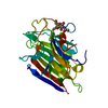
| ||||||||
|---|---|---|---|---|---|---|---|---|---|
| 1 | 
| ||||||||
| Unit cell |
|
-Components
| #1: Protein | Mass: 25610.604 Da / Num. of mol.: 1 / Fragment: RESIDUES 32-268 / Source method: isolated from a natural source / Source: (natural) Robinia pseudoacacia (black locust) / Tissue: BARK / References: UniProt: Q41159 |
|---|---|
| #2: Sugar | ChemComp-A2G /   Type: D-saccharide, alpha linking / Mass: 221.208 Da / Num. of mol.: 1 Type: D-saccharide, alpha linking / Mass: 221.208 Da / Num. of mol.: 1Source method: isolated from a genetically manipulated source Formula: C8H15NO6 |
| #3: Chemical | ChemComp-CA /   Mass: 40.078 Da / Num. of mol.: 1 / Source method: obtained synthetically / Formula: Ca Mass: 40.078 Da / Num. of mol.: 1 / Source method: obtained synthetically / Formula: Ca |
| #4: Water | ChemComp-HOH /  Mass: 18.015 Da / Num. of mol.: 150 / Source method: isolated from a natural source / Formula: H2O Mass: 18.015 Da / Num. of mol.: 150 / Source method: isolated from a natural source / Formula: H2O |
| Sequence details | AMINO ACID HETEROGENE |
-Experimental details
-Experiment
| Experiment | Method: X-RAY DIFFRACTION / Number of used crystals: 1 |
|---|
- Sample preparation
| Crystal | Density Matthews: 2.84 Å3/Da / Density % sol: 56.71 % | |||||||||||||||||||||||||
|---|---|---|---|---|---|---|---|---|---|---|---|---|---|---|---|---|---|---|---|---|---|---|---|---|---|---|
| Crystal grow | Temperature: 277 K / Method: vapor diffusion, hanging drop / pH: 4.8 Details: 0.2M ammonium sulphate, 30% polyethylene glycol 4000, pH 4.8, VAPOR DIFFUSION, HANGING DROP, temperature 277K | |||||||||||||||||||||||||
| Crystal grow | *PLUS pH: 8 Details: Rabijns, A., (2000) Acta Crystallogr., Sect.D, 56, 1638. | |||||||||||||||||||||||||
| Components of the solutions | *PLUS
|
-Data collection
| Diffraction | Mean temperature: 100 K |
|---|---|
| Diffraction source | Source: SYNCHROTRON / Site: ELETTRA  / Beamline: 5.2R / Wavelength: 1 / Beamline: 5.2R / Wavelength: 1 |
| Detector | Type: MARRESEARCH / Detector: IMAGE PLATE / Date: May 22, 2000 |
| Radiation | Protocol: SINGLE WAVELENGTH / Monochromatic (M) / Laue (L): M / Scattering type: x-ray |
| Radiation wavelength | Wavelength: 1 Å / Relative weight: 1 |
| Reflection | Resolution: 2.05→20 Å / Num. all: 18204 / Num. obs: 18204 / % possible obs: 77.2 % / Observed criterion σ(F): 1.41 / Observed criterion σ(I): 2 / Redundancy: 2.94 % / Biso Wilson estimate: 21.748 Å2 / Rmerge(I) obs: 0.086 / Net I/σ(I): 11.87 |
| Reflection shell | Resolution: 2.05→2.09 Å / Redundancy: 2.95 % / Rmerge(I) obs: 0.278 / Num. unique all: 893 / % possible all: 96.5 |
| Reflection | *PLUS Lowest resolution: 20 Å / Observed criterion σ(I): 2 / Num. measured all: 53508 |
- Processing
| Software |
| |||||||||||||||||||||||||
|---|---|---|---|---|---|---|---|---|---|---|---|---|---|---|---|---|---|---|---|---|---|---|---|---|---|---|
| Refinement | Resolution: 2.05→20 Å / σ(F): 0 / σ(I): 0 / Stereochemistry target values: Engh and Huber
| |||||||||||||||||||||||||
| Refinement step | Cycle: LAST / Resolution: 2.05→20 Å
| |||||||||||||||||||||||||
| Refine LS restraints |
| |||||||||||||||||||||||||
| Software | *PLUS Name: CNS / Classification: refinement | |||||||||||||||||||||||||
| Refinement | *PLUS Lowest resolution: 20 Å / σ(F): 0 / Rfactor obs: 0.197 | |||||||||||||||||||||||||
| Solvent computation | *PLUS | |||||||||||||||||||||||||
| Displacement parameters | *PLUS |
