 Movie
Movie Controller


+ Open data

- Basic information
| Entry | Database: PDB / ID: 1les | ||||||||||||
|---|---|---|---|---|---|---|---|---|---|---|---|---|---|
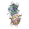
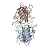
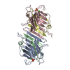
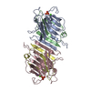
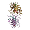
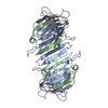
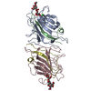
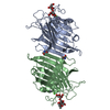
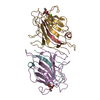
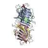
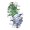
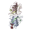
| Title | LENTIL LECTIN COMPLEXED WITH SUCROSE | ||||||||||||
 Components Components | (LENTIL LECTIN) x 2 | ||||||||||||
 Keywords Keywords | LECTIN / PROTEIN-SUGAR INTERACTIONS | ||||||||||||
| Function / homology |  Function and homology information Function and homology informationcarbohydrate mediated signaling / D-mannose binding / manganese ion binding / carbohydrate binding / calcium ion binding Similarity search - Function | ||||||||||||
| Biological species |  Lens culinaris (lentil) Lens culinaris (lentil) | ||||||||||||
| Method |  X-RAY DIFFRACTION / Resolution: 1.9 Å X-RAY DIFFRACTION / Resolution: 1.9 Å | ||||||||||||
 Authors Authors | Hamelryck, T. / Loris, R. | ||||||||||||
 Citation Citation | Journal: J.Biol.Chem. / Year: 1995 Title: NMR, molecular modeling, and crystallographic studies of lentil lectin-sucrose interaction. Authors: Casset, F. / Hamelryck, T. / Loris, R. / Brisson, J.R. / Tellier, C. / Dao-Thi, M.H. / Wyns, L. / Poortmans, F. / Perez, S. / Imberty, A. #1: Journal: J.Mol.Biol. / Year: 1994Title: Conserved Waters in Legume Lectin Crystal Structures: The Importance of Bound Waters for the Sequence-Structure Relationship within the Legume Lectin Family Authors: Loris, R. / Stas, P.P.G. / Wyns, L. #2: Journal: Biochemistry / Year: 1993Title: Crystal Structure Determination and Refinement at 2.3 Angstroms Resolution of the Lentil Lectin Authors: Loris, R. / Steyaert, J. / Maes, D. / Lisgarten, J. / Pickersgill, R. / Wyns, L. #3: Journal: J.Mol.Biol. / Year: 1992Title: Two Crystal Forms of the Lentil Lectin Diffract to High Resolution Authors: Loris, R. / Lisgarten, J. / Maes, D. / Pickersgill, R. / Koerber, F. / Reynolds, C. / Wyns, L. | ||||||||||||
| History |
|
- Structure visualization
| Structure viewer | Molecule: MolmilJmol/JSmol |
|---|
- Downloads & links
-Download
| PDBx/mmCIF format | 1les.cif.gz | 109.7 KB | Display | PDBx/mmCIF format |
|---|---|---|---|---|
| PDB format | pdb1les.ent.gz | 83 KB | Display | PDB format |
| PDBx/mmJSON format | 1les.json.gz | Tree view | PDBx/mmJSON format | |
| Others |  Other downloads Other downloads |
-Validation report
| Arichive directory | https://data.pdbj.org/pub/pdb/validation_reports/le/1lesftp://data.pdbj.org/pub/pdb/validation_reports/le/1les | HTTPS FTP |
|---|
-Related structure data
| Similar structure data |
|---|


-Links
 PDBj
PDBj

- Assembly
| Deposited unit | 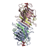
| ||||||||
|---|---|---|---|---|---|---|---|---|---|
| 1 |
| ||||||||
| Unit cell |
| ||||||||
| Atom site foot note | 1: ALA A 80 - ASP A 81 OMEGA = 0.57 PEPTIDE BOND DEVIATES SIGNIFICANTLY FROM TRANS CONFORMATION 2: ALA C 80 - ASP C 81 OMEGA = 0.33 PEPTIDE BOND DEVIATES SIGNIFICANTLY FROM TRANS CONFORMATION 3: GLY B 47 - HIS B 47 OMEGA = 0.02 PEPTIDE BOND DEVIATES SIGNIFICANTLY FROM TRANS CONFORMATION 4: GLY D 47 - HIS D 47 OMEGA = 0.02 PEPTIDE BOND DEVIATES SIGNIFICANTLY FROM TRANS CONFORMATION | ||||||||
| Noncrystallographic symmetry (NCS) | NCS oper: (Code: given Matrix: (0.3718, 0.0008, -0.9283), Vector: Details | THE LENTIL LECTIN MOLECULE NORMALLY EXISTS AS A DIMER. THE TWO MONOMERS IN THIS ENTRY ARE RELATED BY A PSEUDO TWO-FOLD AXIS. EACH MONOMER CONSISTS OF TWO SEPARATE POLYPEPTIDE CHAINS, ALPHA AND BETA. THE ALPHA CHAIN CONSISTS OF 181 RESIDUES AND THE BETA CHAIN CONSISTS OF 52 RESIDUES. THE ALPHA AND BETA CHAINS OF MONOMER 1 HAVE BEEN ASSIGNED CHAIN IDENTIFIERS *A* AND *B*, RESPECTIVELY, IN THIS ENTRY. THE ALPHA AND BETA CHAINS OF MONOMER 2 HAVE BEEN ASSIGNED CHAIN IDENTIFIERS *C* AND *D*, RESPECTIVELY, IN THIS ENTRY. THIS NUMBERING SCHEME IS THE SAME AS THAT USED FOR PEA LECTIN BY EINSPAHR ET AL. (ENTRY 2LTN). THE MONOMERS ARE RELATED BY A NON-CRYSTALLOGRAPHIC TWO-FOLD AXIS PARALLEL TO THE CRYSTALLOGRAPHIC B-AXIS. THE TRANSFORMATION PRESENTED ON *MTRIX* RECORDS BELOW WILL YIELD APPROXIMATE COORDINATES FOR CHAINS *C* AND *D* WHEN APPLIED TO CHAINS *A* AND *B*, RESPECTIVELY. THE RMS BETWEEN THE BACKBONE COORDINATES IS 0.12 ANGSTROMS. | |
-Components
-Protein , 2 types, 4 molecules ACBD
| #1: Protein | Mass: 19906.982 Da / Num. of mol.: 2 / Source method: isolated from a natural source / Source: (natural) Lens culinaris (lentil) / Organ: SEED / References: PIR: LNLWBA, UniProt: P02870*PLUS#2: Protein | Mass: 5748.350 Da / Num. of mol.: 2 / Source method: isolated from a natural source / Source: (natural) Lens culinaris (lentil) / Organ: SEED / References: UniProt: P02870 |
|---|
-Sugars , 1 types, 2 molecules
| #3: Polysaccharide |   Source method: isolated from a genetically manipulated source Details: oligosaccharide with reducing-end-to-reducing-end glycosidic bond References: sucrose |
|---|
-Non-polymers , 3 types, 232 molecules 


| #4: Chemical |  Mass: 40.078 Da / Num. of mol.: 2 / Source method: obtained synthetically / Formula: Ca Mass: 40.078 Da / Num. of mol.: 2 / Source method: obtained synthetically / Formula: Ca#5: Chemical |  Mass: 54.938 Da / Num. of mol.: 2 / Source method: obtained synthetically / Formula: Mn Mass: 54.938 Da / Num. of mol.: 2 / Source method: obtained synthetically / Formula: Mn#6: Water | ChemComp-HOH / | Mass: 18.015 Da / Num. of mol.: 228 / Source method: isolated from a natural source / Formula: H2O |
|---|
-Details
| Nonpolymer details | EACH MONOMER HAS A BOUND CALCIUM, MANGANESE, AND SUCROSE MOLECULE. THE CALCIUM AND MANGANESE IONS ...EACH MONOMER HAS A BOUND CALCIUM, MANGANESE, AND SUCROSE MOLECULE. THE CALCIUM AND MANGANESE IONS ARE ESSENTIAL FOR STABILIZIN |
|---|
-Experimental details
-Experiment
| Experiment | Method: X-RAY DIFFRACTION / Number of used crystals: 1 |
|---|
- Sample preparation
| Crystal | Density Matthews: 2.82 Å3/Da / Density % sol: 56.46 % | |||||||||||||||||||||||||||||||||||
|---|---|---|---|---|---|---|---|---|---|---|---|---|---|---|---|---|---|---|---|---|---|---|---|---|---|---|---|---|---|---|---|---|---|---|---|---|
| Crystal grow | pH: 6.5 / Details: pH 6.5 | |||||||||||||||||||||||||||||||||||
| Crystal grow | *PLUS Method: vapor diffusion, hanging drop | |||||||||||||||||||||||||||||||||||
| Components of the solutions | *PLUS
|
-Data collection
| Diffraction source | Wavelength: 1.5418 Å |
|---|---|
| Detector | Type: ENRAF-NONIUS FAST / Detector: DIFFRACTOMETER / Date: Jun 24, 1994 |
| Radiation | Monochromatic (M) / Laue (L): M / Scattering type: x-ray |
| Radiation wavelength | Wavelength: 1.5418 Å / Relative weight: 1 |
| Reflection | Resolution: 1.79→15 Å / Num. obs: 43108 / % possible obs: 69.9 % / Redundancy: 2.5 % / Rmerge(I) obs: 0.073 |
| Reflection | *PLUS Highest resolution: 1.9 Å / Num. measured all: 114331 / Rmerge(I) obs: 0.073 |
| Reflection shell | *PLUS Highest resolution: 1.9 Å / Lowest resolution: 1.96 Å / % possible obs: 75.1 % / Rmerge(I) obs: 0.295 |
- Processing
| Software |
| ||||||||||||||||||||||||||||||||||||||||||||||||||||||||||||
|---|---|---|---|---|---|---|---|---|---|---|---|---|---|---|---|---|---|---|---|---|---|---|---|---|---|---|---|---|---|---|---|---|---|---|---|---|---|---|---|---|---|---|---|---|---|---|---|---|---|---|---|---|---|---|---|---|---|---|---|---|---|
| Refinement | Resolution: 1.9→6 Å / σ(F): 0 /
| ||||||||||||||||||||||||||||||||||||||||||||||||||||||||||||
| Displacement parameters | Biso mean: 23.51 Å2 | ||||||||||||||||||||||||||||||||||||||||||||||||||||||||||||
| Refine analyze | Luzzati coordinate error obs: 0.2 Å | ||||||||||||||||||||||||||||||||||||||||||||||||||||||||||||
| Refinement step | Cycle: LAST / Resolution: 1.9→6 Å
| ||||||||||||||||||||||||||||||||||||||||||||||||||||||||||||
| Refine LS restraints |
| ||||||||||||||||||||||||||||||||||||||||||||||||||||||||||||
| Software | *PLUS Name: X-PLOR / Classification: refinement | ||||||||||||||||||||||||||||||||||||||||||||||||||||||||||||
| Refinement | *PLUS | ||||||||||||||||||||||||||||||||||||||||||||||||||||||||||||
| Solvent computation | *PLUS | ||||||||||||||||||||||||||||||||||||||||||||||||||||||||||||
| Displacement parameters | *PLUS | ||||||||||||||||||||||||||||||||||||||||||||||||||||||||||||
| Refine LS restraints | *PLUS
|
