 Movie
Movie Controller

[English] 日本語
 Yorodumi
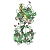
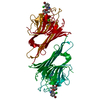
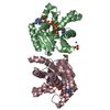
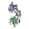
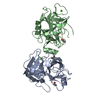
- PDB-4g0d: Human collagenase 3 (MMP-13) full form with peptides from pro-domain -
+ Open data

- Basic information
| Entry | Database: PDB / ID: 4g0d | ||||||
|---|---|---|---|---|---|---|---|
| Title | Human collagenase 3 (MMP-13) full form with peptides from pro-domain | ||||||
 Components Components |
| ||||||
 Keywords Keywords | HYDROLASE / protein-peptide complex / collagenase / cleavage with mmp3 / pro-peptide / metzincin / Zinc metalloprotease / collagen cleavage / collagen | ||||||
| Function / homology |  Function and homology information Function and homology informationgrowth plate cartilage development / RUNX2 regulates genes involved in cell migration / endochondral ossification / Hydrolases; Acting on peptide bonds (peptidases); Metalloendopeptidases / bone morphogenesis / Assembly of collagen fibrils and other multimeric structures / bone mineralization / Activation of Matrix Metalloproteinases / response to amyloid-beta / Collagen degradation ...growth plate cartilage development / RUNX2 regulates genes involved in cell migration / endochondral ossification / Hydrolases; Acting on peptide bonds (peptidases); Metalloendopeptidases / bone morphogenesis / Assembly of collagen fibrils and other multimeric structures / bone mineralization / Activation of Matrix Metalloproteinases / response to amyloid-beta / Collagen degradation / collagen catabolic process / extracellular matrix disassembly / Degradation of the extracellular matrix / collagen binding / extracellular matrix organization / metalloendopeptidase activity / extracellular matrix / endopeptidase activity / serine-type endopeptidase activity / calcium ion binding / proteolysis / : / extracellular region / zinc ion binding Similarity search - Function | ||||||
| Biological species |  Homo sapiens (human) Homo sapiens (human) | ||||||
| Method |  X-RAY DIFFRACTION / SYNCHROTRON / MOLECULAR REPLACEMENT / Resolution: 2.54 Å X-RAY DIFFRACTION / SYNCHROTRON / MOLECULAR REPLACEMENT / Resolution: 2.54 Å | ||||||
 Authors Authors | Stura, E.A. / Vera, L. / Visse, R. / Nagase, H. / Dive, V. | ||||||
 Citation Citation | Journal: Faseb J. / Year: 2013 Title: Crystal structure of full-length human collagenase 3 (MMP-13) with peptides in the active site defines exosites in the catalytic domain. Authors: Stura, E.A. / Visse, R. / Cuniasse, P. / Dive, V. / Nagase, H. | ||||||
| History |
|
- Structure visualization
| Structure viewer | Molecule: MolmilJmol/JSmol |
|---|
- Downloads & links
-Download
| PDBx/mmCIF format | 4g0d.cif.gz | 363.9 KB | Display | PDBx/mmCIF format |
|---|---|---|---|---|
| PDB format | pdb4g0d.ent.gz | 294.5 KB | Display | PDB format |
| PDBx/mmJSON format | 4g0d.json.gz | Tree view | PDBx/mmJSON format | |
| Others |  Other downloads Other downloads |
-Validation report
| Arichive directory | https://data.pdbj.org/pub/pdb/validation_reports/g0/4g0dftp://data.pdbj.org/pub/pdb/validation_reports/g0/4g0d | HTTPS FTP |
|---|
-Related structure data
| Related structure data |  4fu4C  4fvlSC C: citing same article ( S: Starting model for refinement |
|---|---|
| Similar structure data |


-Links
 PDBj
PDBj


- Assembly
| Deposited unit | 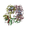
| ||||||||
|---|---|---|---|---|---|---|---|---|---|
| 1 | 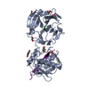
| ||||||||
| 2 | 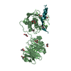
| ||||||||
| 3 | 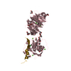
| ||||||||
| 4 | 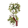
| ||||||||
| Unit cell |
|
-Components
-Protein / Protein/peptide , 2 types, 8 molecules ABCDWXYZ
| #1: Protein | Mass: 42284.617 Da / Num. of mol.: 4 / Fragment: Inactive full form (UNP residues 104-471) / Mutation: E223A Source method: isolated from a genetically manipulated source Source: (gene. exp.) Homo sapiens (human) / Gene: MMP13 / Plasmid: PET3A / Production host:  References: UniProt: P45452, Hydrolases; Acting on peptide bonds (peptidases); Metalloendopeptidases #2: Protein/peptide | Mass: 3109.209 Da / Num. of mol.: 4 / Fragment: pro-domain fragment (UNP residues 25-50) Source method: isolated from a genetically manipulated source Source: (gene. exp.) Homo sapiens (human) / Gene: MMP13 / Plasmid: PET3A / Production host: |
|---|
-Non-polymers , 7 types, 982 molecules 


| #3: Chemical | ChemComp-ZN /  Mass: 65.409 Da / Num. of mol.: 8 / Source method: obtained synthetically / Formula: Zn Mass: 65.409 Da / Num. of mol.: 8 / Source method: obtained synthetically / Formula: Zn#4: Chemical | ChemComp-CA /  Mass: 40.078 Da / Num. of mol.: 23 / Source method: obtained synthetically / Formula: Ca Mass: 40.078 Da / Num. of mol.: 23 / Source method: obtained synthetically / Formula: Ca#5: Chemical | ChemComp-CL /  Mass: 35.453 Da / Num. of mol.: 8 / Source method: obtained synthetically / Formula: Cl Mass: 35.453 Da / Num. of mol.: 8 / Source method: obtained synthetically / Formula: Cl#6: Chemical | ChemComp-PGO /  Mass: 76.094 Da / Num. of mol.: 38 / Source method: obtained synthetically / Formula: C3H8O2 Mass: 76.094 Da / Num. of mol.: 38 / Source method: obtained synthetically / Formula: C3H8O2#7: Chemical | ChemComp-PEG /  Mass: 106.120 Da / Num. of mol.: 4 / Source method: obtained synthetically / Formula: C4H10O3 Mass: 106.120 Da / Num. of mol.: 4 / Source method: obtained synthetically / Formula: C4H10O3#8: Chemical | ChemComp-GOL /  Mass: 92.094 Da / Num. of mol.: 5 / Source method: obtained synthetically / Formula: C3H8O3 Mass: 92.094 Da / Num. of mol.: 5 / Source method: obtained synthetically / Formula: C3H8O3#9: Water | ChemComp-HOH / | Mass: 18.015 Da / Num. of mol.: 896 / Source method: isolated from a natural source / Formula: H2O |
|---|
-Details
| Has protein modification | Y |
|---|---|
| Sequence details | AFTER CLEAVAGE WITH PROTEASE MMP3 TO CUT OFF THE PRO-DOMAIN TO GIVE RISE TO THE MATURE (INACTIVE) ...AFTER CLEAVAGE WITH PROTEASE MMP3 TO CUT OFF THE PRO-DOMAIN TO GIVE RISE TO THE MATURE (INACTIVE) PROTEASE (E223A), A FRAGMENT OF THE PRO-PEPTIDE IS BOUND BACK ONTO THE INACTIVE PROTEASE. THUS CHAIN W BELONGS TO CHAIN A, CHAIN X TO CHAIN B, Y TO C AND Z TO CHAIN D. |
-Experimental details
-Experiment
| Experiment | Method: X-RAY DIFFRACTION / Number of used crystals: 1 |
|---|
- Sample preparation
| Crystal | Density Matthews: 2.92 Å3/Da / Density % sol: 57.89 % |
|---|---|
| Crystal grow | Temperature: 277 K / Method: slow cooling / pH: 7.5 Details: propeptide impurity induces crystallization on cold storage Cryoprotectant: 10% PEG 10K, 5% di-ethylene glycol, 20% 1.2-propanediol, 5% glycerol, .2 M NaCl, 10% PCTP buffer 8:2 ratio, pH 7. ...Details: propeptide impurity induces crystallization on cold storage Cryoprotectant: 10% PEG 10K, 5% di-ethylene glycol, 20% 1.2-propanediol, 5% glycerol, .2 M NaCl, 10% PCTP buffer 8:2 ratio, pH 7.5, SLOW COOLING, temperature 277.0K |
-Data collection
| Diffraction | Mean temperature: 100 K | ||||||||||||||||||||||||||||||||||||||||||||||||||||||||||||||||||||||
|---|---|---|---|---|---|---|---|---|---|---|---|---|---|---|---|---|---|---|---|---|---|---|---|---|---|---|---|---|---|---|---|---|---|---|---|---|---|---|---|---|---|---|---|---|---|---|---|---|---|---|---|---|---|---|---|---|---|---|---|---|---|---|---|---|---|---|---|---|---|---|---|
| Diffraction source | Source: SYNCHROTRON / Site: ESRF  / Beamline: ID23-2 / Wavelength: 0.8726 Å / Beamline: ID23-2 / Wavelength: 0.8726 Å | ||||||||||||||||||||||||||||||||||||||||||||||||||||||||||||||||||||||
| Detector | Type: MAR scanner 345 mm plate / Detector: IMAGE PLATE / Date: Jan 31, 2011 Details: Pt coated mirrors in a Kirkpatrick-Baez (KB) geometry | ||||||||||||||||||||||||||||||||||||||||||||||||||||||||||||||||||||||
| Radiation | Monochromator: SAGITALLY FOCUSED Si(111) / Protocol: SINGLE WAVELENGTH / Monochromatic (M) / Laue (L): M / Scattering type: x-ray | ||||||||||||||||||||||||||||||||||||||||||||||||||||||||||||||||||||||
| Radiation wavelength | Wavelength: 0.8726 Å / Relative weight: 1 | ||||||||||||||||||||||||||||||||||||||||||||||||||||||||||||||||||||||
| Reflection | Resolution: 2.54→50 Å / Num. all: 69045 / Num. obs: 68894 / % possible obs: 99.7 % / Observed criterion σ(F): 0 / Observed criterion σ(I): -3 / Redundancy: 4.05 % / Biso Wilson estimate: 39.978 Å2 / Rmerge(I) obs: 0.258 / Rsym value: 0.224 / Net I/σ(I): 8.47 | ||||||||||||||||||||||||||||||||||||||||||||||||||||||||||||||||||||||
| Reflection shell | Diffraction-ID: 1
|
- Processing
| Software |
| ||||||||||||||||||||||||||||||||||||||||||||||||||||||||||||||||||||||||||||||||||||||||||||||||||||||||||||||||||||||||||||||||||||||||||||||||||||||||||||||||||||||||||||||||||||||
|---|---|---|---|---|---|---|---|---|---|---|---|---|---|---|---|---|---|---|---|---|---|---|---|---|---|---|---|---|---|---|---|---|---|---|---|---|---|---|---|---|---|---|---|---|---|---|---|---|---|---|---|---|---|---|---|---|---|---|---|---|---|---|---|---|---|---|---|---|---|---|---|---|---|---|---|---|---|---|---|---|---|---|---|---|---|---|---|---|---|---|---|---|---|---|---|---|---|---|---|---|---|---|---|---|---|---|---|---|---|---|---|---|---|---|---|---|---|---|---|---|---|---|---|---|---|---|---|---|---|---|---|---|---|---|---|---|---|---|---|---|---|---|---|---|---|---|---|---|---|---|---|---|---|---|---|---|---|---|---|---|---|---|---|---|---|---|---|---|---|---|---|---|---|---|---|---|---|---|---|---|---|---|---|
| Refinement | Method to determine structure: MOLECULAR REPLACEMENT Starting model: 4FVL Resolution: 2.54→49.503 Å / SU ML: 0.37 / Isotropic thermal model: Isotropic / Cross valid method: THROUGHOUT / σ(F): 1.99 / σ(I): -3 / Phase error: 25.11 / Stereochemistry target values: ML
| ||||||||||||||||||||||||||||||||||||||||||||||||||||||||||||||||||||||||||||||||||||||||||||||||||||||||||||||||||||||||||||||||||||||||||||||||||||||||||||||||||||||||||||||||||||||
| Solvent computation | Shrinkage radii: 0.9 Å / VDW probe radii: 1.11 Å / Solvent model: FLAT BULK SOLVENT MODEL | ||||||||||||||||||||||||||||||||||||||||||||||||||||||||||||||||||||||||||||||||||||||||||||||||||||||||||||||||||||||||||||||||||||||||||||||||||||||||||||||||||||||||||||||||||||||
| Displacement parameters | Biso mean: 28.877 Å2 | ||||||||||||||||||||||||||||||||||||||||||||||||||||||||||||||||||||||||||||||||||||||||||||||||||||||||||||||||||||||||||||||||||||||||||||||||||||||||||||||||||||||||||||||||||||||
| Refinement step | Cycle: LAST / Resolution: 2.54→49.503 Å
| ||||||||||||||||||||||||||||||||||||||||||||||||||||||||||||||||||||||||||||||||||||||||||||||||||||||||||||||||||||||||||||||||||||||||||||||||||||||||||||||||||||||||||||||||||||||
| Refine LS restraints |
| ||||||||||||||||||||||||||||||||||||||||||||||||||||||||||||||||||||||||||||||||||||||||||||||||||||||||||||||||||||||||||||||||||||||||||||||||||||||||||||||||||||||||||||||||||||||
| LS refinement shell | Refine-ID: X-RAY DIFFRACTION
|
