 Movie
Movie Controller

[English] 日本語
 Yorodumi
- PDB-5xev: Crystal Structure of a novel Xaa-Pro dipeptidase from Deinococcus... -
+ Open data

- Basic information
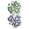
| Entry | Database: PDB / ID: 5xev | ||||||
|---|---|---|---|---|---|---|---|
| Title | Crystal Structure of a novel Xaa-Pro dipeptidase from Deinococcus radiodurans | ||||||
 Components Components | Xaa-Pro dipeptidase,Peptidase-related protein | ||||||
 Keywords Keywords | HYDROLASE / Xaa-Pro dipeptidase / prolidase / M28B peptidase / Deinococcus radiodurans | ||||||
| Function / homology | : / Peptidase M24 / Metallopeptidase family M24 / Creatinase/aminopeptidase-like / metal ion binding / ACETATE ION / PHOSPHATE ION / Peptidase-related protein Function and homology information Function and homology information | ||||||
| Biological species |  Deinococcus radiodurans R1 (radioresistant) Deinococcus radiodurans R1 (radioresistant) | ||||||
| Method |  X-RAY DIFFRACTION / SYNCHROTRON / MOLECULAR REPLACEMENT / Resolution: 1.4 Å X-RAY DIFFRACTION / SYNCHROTRON / MOLECULAR REPLACEMENT / Resolution: 1.4 Å | ||||||
 Authors Authors | Are, V.N. / Kumar, A. / Ghosh, B. / Makde, R.D. | ||||||
 Citation Citation | Journal: Proteins / Year: 2017 Title: Crystal structure of a novel prolidase from Deinococcus radiodurans identifies new subfamily of bacterial prolidases. Authors: Are, V.N. / Jamdar, S.N. / Ghosh, B. / Goyal, V.D. / Kumar, A. / Neema, S. / Gadre, R. / Makde, R.D. | ||||||
| History |
|
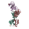
- Structure visualization
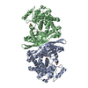
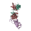
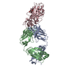
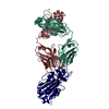
| Structure viewer | Molecule: MolmilJmol/JSmol |
|---|
- Downloads & links
-Download
| PDBx/mmCIF format | 5xev.cif.gz | 251.3 KB | Display | PDBx/mmCIF format |
|---|---|---|---|---|
| PDB format | pdb5xev.ent.gz | 204.8 KB | Display | PDB format |
| PDBx/mmJSON format | 5xev.json.gz | Tree view | PDBx/mmJSON format | |
| Others |  Other downloads Other downloads |
-Validation report
| Arichive directory | https://data.pdbj.org/pub/pdb/validation_reports/xe/5xevftp://data.pdbj.org/pub/pdb/validation_reports/xe/5xev | HTTPS FTP |
|---|
-Related structure data
| Similar structure data |
|---|


-Links
 PDBj
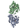
PDBj- Assembly
| Deposited unit | 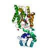
| |||||||||||||||
|---|---|---|---|---|---|---|---|---|---|---|---|---|---|---|---|---|
| 1 | 
| |||||||||||||||
| Unit cell |
| |||||||||||||||
| Components on special symmetry positions |
|
-Components
-Protein , 1 types, 1 molecules A
| #1: Protein | Mass: 45308.289 Da / Num. of mol.: 1 Source method: isolated from a genetically manipulated source Source: (gene. exp.) Deinococcus radiodurans R1 (radioresistant)Plasmid: pST50STRHIS Strain: ATCC 13939 / DSM 20539 / JCM 16871 / LMG 4051 / NBRC 15346 / NCIMB 9279 / R1 / VKM B-1422 Gene: DR_0478 / Details (production host): T7 based expression plasmid / Production host: |
|---|
-Non-polymers , 6 types, 423 molecules 


| #2: Chemical |  Mass: 65.409 Da / Num. of mol.: 2 / Source method: obtained synthetically / Formula: Zn Mass: 65.409 Da / Num. of mol.: 2 / Source method: obtained synthetically / Formula: Zn#3: Chemical | ChemComp-PO4 / |  Mass: 94.971 Da / Num. of mol.: 1 / Source method: obtained synthetically / Formula: PO4 Mass: 94.971 Da / Num. of mol.: 1 / Source method: obtained synthetically / Formula: PO4#4: Chemical | ChemComp-ACT / |  Mass: 59.044 Da / Num. of mol.: 1 / Source method: obtained synthetically / Formula: C2H3O2 Mass: 59.044 Da / Num. of mol.: 1 / Source method: obtained synthetically / Formula: C2H3O2#5: Chemical | ChemComp-CL / |  Mass: 35.453 Da / Num. of mol.: 1 / Source method: obtained synthetically / Formula: Cl Mass: 35.453 Da / Num. of mol.: 1 / Source method: obtained synthetically / Formula: Cl#6: Chemical | ChemComp-EDO / |  Mass: 62.068 Da / Num. of mol.: 1 / Source method: obtained synthetically / Formula: C2H6O2 Mass: 62.068 Da / Num. of mol.: 1 / Source method: obtained synthetically / Formula: C2H6O2#7: Water | ChemComp-HOH / | Mass: 18.015 Da / Num. of mol.: 417 / Source method: isolated from a natural source / Formula: H2O |
|---|
-Details
| Sequence details | This protein corresponds to database(UniParc:UPI0002D66B97,GenBank:ANC72273). |
|---|
-Experimental details
-Experiment
| Experiment | Method: X-RAY DIFFRACTION / Number of used crystals: 1 |
|---|
- Sample preparation
| Crystal | Density Matthews: 2.45 Å3/Da / Density % sol: 49.72 % |
|---|---|
| Crystal grow | Temperature: 294 K / Method: microbatch / pH: 4.8 Details: 40 mM KH2PO4, 10mM ammonium citrate, pH 4.8, 3 mM phenylalanine, 1 mM MnCl2, 10 mM ZnCl2, 16% PEG8000, 20% Glycerol PH range: 4.5-5.8 |
-Data collection
| Diffraction | Mean temperature: 100 K |
|---|---|
| Diffraction source | Source: SYNCHROTRON / Site: RRCAT INDUS-2  / Beamline: PX-BL21 / Wavelength: 0.97947 Å / Beamline: PX-BL21 / Wavelength: 0.97947 Å |
| Detector | Type: MARMOSAIC 225 mm CCD / Detector: CCD / Date: Dec 29, 2016 / Details: Mirrors |
| Radiation | Monochromator: Si111 / Protocol: SINGLE WAVELENGTH / Monochromatic (M) / Laue (L): M / Scattering type: x-ray |
| Radiation wavelength | Wavelength: 0.97947 Å / Relative weight: 1 |
| Reflection | Resolution: 1.4→45.94 Å / Num. obs: 85429 / % possible obs: 99.9 % / Redundancy: 6.7 % / CC1/2: 0.999 / Rmerge(I) obs: 0.066 / Rpim(I) all: 0.027 / Net I/σ(I): 15.9 |
| Reflection shell | Resolution: 1.4→1.42 Å / Redundancy: 6.6 % / Rmerge(I) obs: 0.789 / Mean I/σ(I) obs: 2.3 / Num. unique obs: 4196 / CC1/2: 0.757 / Rpim(I) all: 0.331 / % possible all: 99.4 |
- Processing
| Software |
| |||||||||||||||||||||||||||||||||||||||||||||||||||||||||||||||||||||||||||||||||||||||||||||||||||||||||||||||||||||||||||||||||||||||||||||||||||||||||||||||||||||||||||||||||||||||||||||||||||||||||||||||||||||||||
|---|---|---|---|---|---|---|---|---|---|---|---|---|---|---|---|---|---|---|---|---|---|---|---|---|---|---|---|---|---|---|---|---|---|---|---|---|---|---|---|---|---|---|---|---|---|---|---|---|---|---|---|---|---|---|---|---|---|---|---|---|---|---|---|---|---|---|---|---|---|---|---|---|---|---|---|---|---|---|---|---|---|---|---|---|---|---|---|---|---|---|---|---|---|---|---|---|---|---|---|---|---|---|---|---|---|---|---|---|---|---|---|---|---|---|---|---|---|---|---|---|---|---|---|---|---|---|---|---|---|---|---|---|---|---|---|---|---|---|---|---|---|---|---|---|---|---|---|---|---|---|---|---|---|---|---|---|---|---|---|---|---|---|---|---|---|---|---|---|---|---|---|---|---|---|---|---|---|---|---|---|---|---|---|---|---|---|---|---|---|---|---|---|---|---|---|---|---|---|---|---|---|---|---|---|---|---|---|---|---|---|---|---|---|---|---|---|---|---|
| Refinement | Method to determine structure: MOLECULAR REPLACEMENT / Resolution: 1.4→31.955 Å / SU ML: 0.12 / Cross valid method: FREE R-VALUE / σ(F): 1.35 / Phase error: 15.77
| |||||||||||||||||||||||||||||||||||||||||||||||||||||||||||||||||||||||||||||||||||||||||||||||||||||||||||||||||||||||||||||||||||||||||||||||||||||||||||||||||||||||||||||||||||||||||||||||||||||||||||||||||||||||||
| Solvent computation | Shrinkage radii: 0.9 Å / VDW probe radii: 1.11 Å | |||||||||||||||||||||||||||||||||||||||||||||||||||||||||||||||||||||||||||||||||||||||||||||||||||||||||||||||||||||||||||||||||||||||||||||||||||||||||||||||||||||||||||||||||||||||||||||||||||||||||||||||||||||||||
| Refinement step | Cycle: LAST / Resolution: 1.4→31.955 Å
| |||||||||||||||||||||||||||||||||||||||||||||||||||||||||||||||||||||||||||||||||||||||||||||||||||||||||||||||||||||||||||||||||||||||||||||||||||||||||||||||||||||||||||||||||||||||||||||||||||||||||||||||||||||||||
| Refine LS restraints |
| |||||||||||||||||||||||||||||||||||||||||||||||||||||||||||||||||||||||||||||||||||||||||||||||||||||||||||||||||||||||||||||||||||||||||||||||||||||||||||||||||||||||||||||||||||||||||||||||||||||||||||||||||||||||||
| LS refinement shell |
| |||||||||||||||||||||||||||||||||||||||||||||||||||||||||||||||||||||||||||||||||||||||||||||||||||||||||||||||||||||||||||||||||||||||||||||||||||||||||||||||||||||||||||||||||||||||||||||||||||||||||||||||||||||||||
| Refinement TLS params. | Method: refined / Origin x: -21.9916 Å / Origin y: -21.9256 Å / Origin z: 1.6981 Å
| |||||||||||||||||||||||||||||||||||||||||||||||||||||||||||||||||||||||||||||||||||||||||||||||||||||||||||||||||||||||||||||||||||||||||||||||||||||||||||||||||||||||||||||||||||||||||||||||||||||||||||||||||||||||||
| Refinement TLS group | Selection details: all |
