 Movie
Movie Controller

[English] 日本語
 Yorodumi
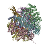
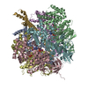
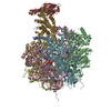
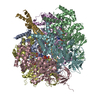
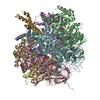
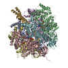
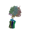
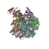
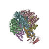
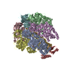
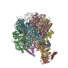
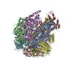
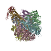
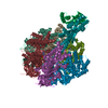
- PDB-1h8e: (ADP.AlF4)2(ADP.SO4) bovine F1-ATPase (all three catalytic sites ... -
+ Open data

- Basic information
| Entry | Database: PDB / ID: 1h8e | ||||||
|---|---|---|---|---|---|---|---|
| Title | (ADP.AlF4)2(ADP.SO4) bovine F1-ATPase (all three catalytic sites occupied) | ||||||
 Components Components | (BOVINE MITOCHONDRIAL F1- ...) x 5 | ||||||
 Keywords Keywords | HYDROLASE / ATP PHOSPHORYLASE / ATP PHOSPHORYLASE (H+ TRANSPORTING) / ATP SYNTHASE / F1FO ATP SYNTHASE / F1-ATPASE | ||||||
| Function / homology |  Function and homology information Function and homology informationFormation of ATP by chemiosmotic coupling / Cristae formation / mitochondrial proton-transporting ATP synthase complex assembly / mitochondrial envelope / Mitochondrial protein degradation / proton transmembrane transporter activity / proton motive force-driven ATP synthesis / proton motive force-driven mitochondrial ATP synthesis / H+-transporting two-sector ATPase / proton-transporting ATP synthase complex ...Formation of ATP by chemiosmotic coupling / Cristae formation / mitochondrial proton-transporting ATP synthase complex assembly / mitochondrial envelope / Mitochondrial protein degradation / proton transmembrane transporter activity / proton motive force-driven ATP synthesis / proton motive force-driven mitochondrial ATP synthesis / H+-transporting two-sector ATPase / proton-transporting ATP synthase complex / proton-transporting ATP synthase activity, rotational mechanism / proton transmembrane transport / aerobic respiration / ADP binding / mitochondrial inner membrane / structural molecule activity / ATP hydrolysis activity / mitochondrion / ATP binding / metal ion binding / plasma membrane Similarity search - Function | ||||||
| Biological species |  | ||||||
| Method |  X-RAY DIFFRACTION / SYNCHROTRON / MOLECULAR REPLACEMENT / Resolution: 2 Å X-RAY DIFFRACTION / SYNCHROTRON / MOLECULAR REPLACEMENT / Resolution: 2 Å | ||||||
 Authors Authors | Menz, R.I. / Walker, J.E. / Leslie, A.G.W. | ||||||
 Citation Citation | Journal: Cell(Cambridge,Mass.) / Year: 2001 Title: Structure of Bovine Mitochondrial F1-ATPase with Nucleotide Bound to All Three Catalytic Sites: Implications for the Mechanism of Rotary Catalysis Authors: Menz, R.I. / Walker, J.E. / Leslie, A.G.W. #1: Journal: Nat.Struct.Biol. / Year: 2000Title: The Structure of the Central Stalk in Bovine F1-ATPase at 2.4A Resolution Authors: Gibbons, C. / Montgomery, M.G. / Leslie, A.G.W. / Walker, J.E. #2: Journal: Science / Year: 1999Title: Molecular Architecture of the Rotary Motor in ATP Synthase Authors: Stock, D. / Leslie, A.G.W. / Walker, J.E. #3: Journal: Angew.Chem.Int.Ed.Engl. / Year: 1998Title: ATP Synthesis by Rotary Catalysis (Nobel Lecture) Authors: Walker, J.E. #4: Journal: Nature / Year: 1994Title: Structure at 2.8 A Resolution of F1-ATPase from Bovine Heart Mitochondria Authors: Abrahams, J.P. / Leslie, A.G.W. / Lutter, R. / Walker, J.E. #5: Journal: J.Mol.Biol. / Year: 1993 Title: Crystallization of F1-ATPase from Bovine Heart Mitochondria Authors: Lutter, R. / Abrahams, J.P. / Van Raaij, M.J. / Todd, R.J. / Lundqvist, T. / Buchanan, S.K. / Leslie, A.G. / Walker, J.E. | ||||||
| History |
|
- Structure visualization
| Structure viewer | Molecule: MolmilJmol/JSmol |
|---|
- Downloads & links
-Download
| PDBx/mmCIF format | 1h8e.cif.gz | 656.4 KB | Display | PDBx/mmCIF format |
|---|---|---|---|---|
| PDB format | pdb1h8e.ent.gz | 530.1 KB | Display | PDB format |
| PDBx/mmJSON format | 1h8e.json.gz | Tree view | PDBx/mmJSON format | |
| Others |  Other downloads Other downloads |
-Validation report
| Arichive directory | https://data.pdbj.org/pub/pdb/validation_reports/h8/1h8eftp://data.pdbj.org/pub/pdb/validation_reports/h8/1h8e | HTTPS FTP |
|---|
-Related structure data
| Related structure data | 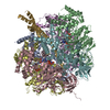 1bmfS S: Starting model for refinement |
|---|---|
| Similar structure data |


-Links
 PDBj
PDBj


- Assembly
| Deposited unit | 
| ||||||||
|---|---|---|---|---|---|---|---|---|---|
| 1 |
| ||||||||
| Unit cell |
| ||||||||
| Details | THE F1-ATPASE MOLECULE HAS THREE COPIES OF THE NON-CATALYTICALPHA SUBUNIT AND THREE COPIES OF THE CATALYTIC BETA SUBUNIT.IN THE 1994 REFERENCE, THE BETA SUBUNITS WERE LABELED ACCORDINGTO THE BOUND NUCLEOTIDE, ASBETA(DP) (BINDS ADP),BETA(E) (NO BOUND NUCLEOTIDE) ANDBETA(TP ) (AMPPNP BOUND).THE ALPHA SUBUNITS (WHICH ALL BIND AMPPNP) CONTRIBUTE TO THECATALYTIC SITES OF THE BETA SUBUNITS, AND HAVE BEEN LABELEDACCORDINGLY . THUSALPHA(DP) CONTRIBUTES TO THE CATALYTIC SITE ON BETA(DP),ALPHA(TP) TO THE SITE ON BETA ( TP) ANDALPHA(E) TO THE SITE ON BETA(E).THE CORRESPONDENCE BETWEEN THE SUBUNIT NAMES AND THE CHAINIDENTIFIERS IS GIVEN BELOW:.CHAIN A: ALPHA(E )CHAIN B: ALPHA(TP)CHAIN C: ALPHA(DP)CHAIN D : BETA(DP)CHAIN E: BETA(E)CHAIN F: BETA(TP) CHAIN G: GAMMA SUBUNITCHAIN H: DELTA SUBUNITCHAIN I: EPSILON SUBUNIT |
-Components
-BOVINE MITOCHONDRIAL F1- ... , 5 types, 9 molecules ABCDEFGHI
| #1: Protein | Mass: 55301.207 Da / Num. of mol.: 3 / Source method: isolated from a natural source / Source: (natural) #2: Protein | Mass: 51757.836 Da / Num. of mol.: 3 / Source method: isolated from a natural source / Source: (natural) #3: Protein | | Mass: 30185.674 Da / Num. of mol.: 1 / Source method: isolated from a natural source / Source: (natural) #4: Protein | | Mass: 15074.813 Da / Num. of mol.: 1 / Source method: isolated from a natural source / Source: (natural) #5: Protein/peptide | | Mass: 5662.693 Da / Num. of mol.: 1 / Source method: isolated from a natural source / Source: (natural) |
|---|
-Non-polymers , 6 types, 1712 molecules 


| #6: Chemical | ChemComp-ADP /  Mass: 427.201 Da / Num. of mol.: 6 / Source method: obtained synthetically / Formula: C10H15N5O10P2 / Comment: ADP, energy-carrying molecule*YM Mass: 427.201 Da / Num. of mol.: 6 / Source method: obtained synthetically / Formula: C10H15N5O10P2 / Comment: ADP, energy-carrying molecule*YM#7: Chemical | ChemComp-MG /  Mass: 24.305 Da / Num. of mol.: 6 / Source method: obtained synthetically / Formula: Mg Mass: 24.305 Da / Num. of mol.: 6 / Source method: obtained synthetically / Formula: Mg#8: Chemical | ChemComp-GOL /  Mass: 92.094 Da / Num. of mol.: 4 / Source method: obtained synthetically / Formula: C3H8O3 Mass: 92.094 Da / Num. of mol.: 4 / Source method: obtained synthetically / Formula: C3H8O3#9: Chemical |  Mass: 102.975 Da / Num. of mol.: 2 / Source method: obtained synthetically / Formula: AlF4 Mass: 102.975 Da / Num. of mol.: 2 / Source method: obtained synthetically / Formula: AlF4#10: Chemical | ChemComp-SO4 / |  Mass: 96.063 Da / Num. of mol.: 1 / Source method: obtained synthetically / Formula: SO4 Mass: 96.063 Da / Num. of mol.: 1 / Source method: obtained synthetically / Formula: SO4#11: Water | ChemComp-HOH / | Mass: 18.015 Da / Num. of mol.: 1693 / Source method: isolated from a natural source / Formula: H2O |
|---|
-Details
| Sequence details | REFERENCE: 1) FOR THE ALPHA SUBUNIT: J. E. WALKER, S. J. POWELL, O. VINAS AND M. J. RUNSWICK, ...REFERENCE: 1) FOR THE ALPHA SUBUNIT: J. E. WALKER, S. J. POWELL, O. VINAS AND M. J. RUNSWICK, BIOCHEMIST |
|---|
-Experimental details
-Experiment
| Experiment | Method: X-RAY DIFFRACTION / Number of used crystals: 1 |
|---|
- Sample preparation
| Crystal | Density Matthews: 2.64 Å3/Da / Density % sol: 54 % | ||||||||||||||||||||||||||||||||||||||||||||||||||||||||||||||||||||||||||||||||||||||||||||||||||||||||||||||||
|---|---|---|---|---|---|---|---|---|---|---|---|---|---|---|---|---|---|---|---|---|---|---|---|---|---|---|---|---|---|---|---|---|---|---|---|---|---|---|---|---|---|---|---|---|---|---|---|---|---|---|---|---|---|---|---|---|---|---|---|---|---|---|---|---|---|---|---|---|---|---|---|---|---|---|---|---|---|---|---|---|---|---|---|---|---|---|---|---|---|---|---|---|---|---|---|---|---|---|---|---|---|---|---|---|---|---|---|---|---|---|---|---|---|
| Crystal grow | pH: 8 Details: CRYSTALS WERE GROWN IN THE PRESENCE OF AZIDE, A KNOWN INHIBITOR, BUT THIS HAS NOT BEEN LOCATED IN THE STRUCTURE., pH 8.00 | ||||||||||||||||||||||||||||||||||||||||||||||||||||||||||||||||||||||||||||||||||||||||||||||||||||||||||||||||
| Crystal grow | *PLUS Temperature: 22-24 ℃ / pH: 7.2 / Method: microdialysis / Details: Lutter, R., (1993) J.Mol.Biol., 229, 787. | ||||||||||||||||||||||||||||||||||||||||||||||||||||||||||||||||||||||||||||||||||||||||||||||||||||||||||||||||
| Components of the solutions | *PLUS
|
-Data collection
| Diffraction | Mean temperature: 100 K |
|---|---|
| Diffraction source | Source: SYNCHROTRON / Site: SRS  / Beamline: PX9.6 / Wavelength: 0.87 / Beamline: PX9.6 / Wavelength: 0.87 |
| Detector | Type: ADSC CCD / Detector: CCD / Date: Aug 15, 1998 |
| Radiation | Protocol: SINGLE WAVELENGTH / Monochromatic (M) / Laue (L): M / Scattering type: x-ray |
| Radiation wavelength | Wavelength: 0.87 Å / Relative weight: 1 |
| Reflection | Resolution: 2→13.5 Å / Num. obs: 209953 / % possible obs: 79.6 % / Observed criterion σ(I): 0 / Redundancy: 2.1 % / Biso Wilson estimate: 26.7 Å2 / Rmerge(I) obs: 0.066 / Net I/σ(I): 9.1 |
| Reflection shell | Resolution: 2→2.14 Å / Redundancy: 1.5 % / Rmerge(I) obs: 0.48 / Mean I/σ(I) obs: 1.7 / % possible all: 40.6 |
| Reflection shell | *PLUS % possible obs: 40.6 % / Rmerge(I) obs: 0.481 / Mean I/σ(I) obs: 1.8 |
- Processing
| Software |
| ||||||||||||||||||||||||||||||||||||||||||||||||||||||||||||||||||||||||||||||||||||
|---|---|---|---|---|---|---|---|---|---|---|---|---|---|---|---|---|---|---|---|---|---|---|---|---|---|---|---|---|---|---|---|---|---|---|---|---|---|---|---|---|---|---|---|---|---|---|---|---|---|---|---|---|---|---|---|---|---|---|---|---|---|---|---|---|---|---|---|---|---|---|---|---|---|---|---|---|---|---|---|---|---|---|---|---|---|
| Refinement | Method to determine structure: MOLECULAR REPLACEMENT Starting model: PDB CODE 1BMF, NATIVE BOVINE MITOCHONDRIAL F1- ATPASE Resolution: 2→13.5 Å / SU B: 5.8 / SU ML: 0.15 / Cross valid method: THROUGHOUT / σ(F): 0 / ESU R: 0.23 / ESU R Free: 0.21 Details: INITIAL REFINEMENT CARRIED OUT WITH TNT. ASP 270 IN CHAINS A, B, C AND THE PEPTIDE BOND BETWEEN ASP 256 AND ASN 257 IN CHAINS D, E, AND F HAVE BEEN MODELED IN A CIS CONFORMATION.
| ||||||||||||||||||||||||||||||||||||||||||||||||||||||||||||||||||||||||||||||||||||
| Displacement parameters | Biso mean: 45 Å2 | ||||||||||||||||||||||||||||||||||||||||||||||||||||||||||||||||||||||||||||||||||||
| Refinement step | Cycle: LAST / Resolution: 2→13.5 Å
| ||||||||||||||||||||||||||||||||||||||||||||||||||||||||||||||||||||||||||||||||||||
| Refine LS restraints |
| ||||||||||||||||||||||||||||||||||||||||||||||||||||||||||||||||||||||||||||||||||||
| Software | *PLUS Name: REFMAC / Classification: refinement | ||||||||||||||||||||||||||||||||||||||||||||||||||||||||||||||||||||||||||||||||||||
| Refinement | *PLUS Rfactor obs: 0.201 | ||||||||||||||||||||||||||||||||||||||||||||||||||||||||||||||||||||||||||||||||||||
| Solvent computation | *PLUS | ||||||||||||||||||||||||||||||||||||||||||||||||||||||||||||||||||||||||||||||||||||
| Displacement parameters | *PLUS |
