 Movie
Movie Controller

[English] 日本語
 Yorodumi
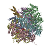
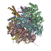
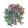
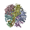
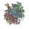
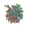
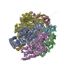
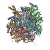
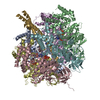
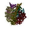
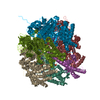
- PDB-1efr: BOVINE MITOCHONDRIAL F1-ATPASE COMPLEXED WITH THE PEPTIDE ANTIBIO... -
+ Open data

- Basic information
| Entry | Database: PDB / ID: 1efr | ||||||
|---|---|---|---|---|---|---|---|
| Title | BOVINE MITOCHONDRIAL F1-ATPASE COMPLEXED WITH THE PEPTIDE ANTIBIOTIC EFRAPEPTIN | ||||||
 Components Components |
| ||||||
 Keywords Keywords | HYDROLASE/ANTIBIOTIC / ANTIBIOTIC / ATP PHOSPHORYLASE / HYDROGEN ION TRANSPORT / ATP SYNTHASE / F1-ATPASE / IONOPHORE / HYDROLASE-ANTIBIOTIC COMPLEX / EFRAPEPTIN / F1-ATPASE-ANTIBIOTIC COMPLEX | ||||||
| Function / homology |  Function and homology information Function and homology informationFormation of ATP by chemiosmotic coupling / Cristae formation / Mitochondrial protein degradation / proton motive force-driven ATP synthesis / proton motive force-driven mitochondrial ATP synthesis / H+-transporting two-sector ATPase / proton-transporting ATP synthase complex / proton-transporting ATP synthase activity, rotational mechanism / ADP binding / mitochondrial inner membrane ...Formation of ATP by chemiosmotic coupling / Cristae formation / Mitochondrial protein degradation / proton motive force-driven ATP synthesis / proton motive force-driven mitochondrial ATP synthesis / H+-transporting two-sector ATPase / proton-transporting ATP synthase complex / proton-transporting ATP synthase activity, rotational mechanism / ADP binding / mitochondrial inner membrane / ATP hydrolysis activity / mitochondrion / ATP binding / metal ion binding / plasma membrane Similarity search - Function | ||||||
| Biological species |  TOLYPOCLADIUM INFLATUM (fungus) TOLYPOCLADIUM INFLATUM (fungus) | ||||||
| Method |  X-RAY DIFFRACTION / SYNCHROTRON / MOLECULAR REPLACEMENT / Resolution: 3.1 Å X-RAY DIFFRACTION / SYNCHROTRON / MOLECULAR REPLACEMENT / Resolution: 3.1 Å | ||||||
 Authors Authors | Abrahams, J.P. / Buchanan, S.K. / Van Raaij, M.J. / Fearnley, I.M. / Leslie, A.G.W. / Walker, J.E. | ||||||
 Citation Citation | Journal: Proc.Natl.Acad.Sci.USA / Year: 1996 Title: The Structure of Bovine F1-ATPase Complexed with the Peptide Antibiotic Efrapeptin. Authors: Abrahams, J.P. / Buchanan, S.K. / Van Raaij, M.J. / Fearnley, I.M. / Leslie, A.G. / Walker, J.E. #1: Journal: Proc.Natl.Acad.Sci.USA / Year: 1996Title: The Structure of Bovine F1-ATPase Complexed with the Antibiotic Inhibitor Aurovertin B Authors: Van Raaij, M.J. / Abrahams, J.P. / Leslie, A.G. / Walker, J.E. #2: Journal: Nature / Year: 1994Title: Structure at 2.8 A Resolution of F1-ATPase from Bovine Heart Mitochondria Authors: Abrahams, J.P. / Leslie, A.G. / Lutter, R. / Walker, J.E. #3: Journal: J.Mol.Biol. / Year: 1993 Title: Crystallization of F1-ATPase from Bovine Heart Mitochondria Authors: Lutter, R. / Abrahams, J.P. / Van Raaij, M.J. / Todd, R.J. / Lundqvist, T. / Buchanan, S.K. / Leslie, A.G. / Walker, J.E. #4: Journal: Embo J. / Year: 1993 Title: Inherent Asymmetry of the Structure of F1-ATPase from Bovine Heart Mitochondria at 6.5 A Resolution Authors: Abrahams, J.P. / Lutter, R. / Todd, R.J. / Van Raaij, M.J. / Leslie, A.G. / Walker, J.E. | ||||||
| History |
| ||||||
| Remark 700 | SHEET DETERMINATION METHOD: DSSP THE SHEETS PRESENTED AS "AA" IN EACH CHAIN ON SHEET RECORDS BELOW ... SHEET DETERMINATION METHOD: DSSP THE SHEETS PRESENTED AS "AA" IN EACH CHAIN ON SHEET RECORDS BELOW IS ACTUALLY AN 10-STRANDED BARREL THIS IS REPRESENTED BY A 11-STRANDED SHEET IN WHICH THE FIRST AND LAST STRANDS ARE IDENTICAL. SHEET DETERMINATION METHOD: DSSP THE SHEETS PRESENTED AS "BA" IN EACH CHAIN ON SHEET RECORDS BELOW IS ACTUALLY AN 10-STRANDED BARREL THIS IS REPRESENTED BY A 11-STRANDED SHEET IN WHICH THE FIRST AND LAST STRANDS ARE IDENTICAL. SHEET DETERMINATION METHOD: DSSP THE SHEETS PRESENTED AS "CA" IN EACH CHAIN ON SHEET RECORDS BELOW IS ACTUALLY AN 10-STRANDED BARREL THIS IS REPRESENTED BY A 11-STRANDED SHEET IN WHICH THE FIRST AND LAST STRANDS ARE IDENTICAL. |
- Structure visualization
| Structure viewer | Molecule: MolmilJmol/JSmol |
|---|
- Downloads & links
-Download
| PDBx/mmCIF format | 1efr.cif.gz | 584.3 KB | Display | PDBx/mmCIF format |
|---|---|---|---|---|
| PDB format | pdb1efr.ent.gz | 477.5 KB | Display | PDB format |
| PDBx/mmJSON format | 1efr.json.gz | Tree view | PDBx/mmJSON format | |
| Others |  Other downloads Other downloads |
-Validation report
| Arichive directory | https://data.pdbj.org/pub/pdb/validation_reports/ef/1efrftp://data.pdbj.org/pub/pdb/validation_reports/ef/1efr | HTTPS FTP |
|---|
-Related structure data
| Related structure data |  1bmfS S: Starting model for refinement |
|---|---|
| Similar structure data |
-Links
 PDBj
PDBj


- Assembly
| Deposited unit | 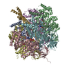
| ||||||||
|---|---|---|---|---|---|---|---|---|---|
| 1 |
| ||||||||
| Unit cell |
|
-Components
-BOVINE MITOCHONDRIAL F1-ATPASE SUBUNIT ... , 3 types, 7 molecules ABCDEFG
| #1: Protein | Mass: 55302.191 Da / Num. of mol.: 3 / Source method: isolated from a natural source / Source: (natural) #2: Protein | Mass: 51757.836 Da / Num. of mol.: 3 / Source method: isolated from a natural source / Source: (natural) #3: Protein | | Mass: 30185.674 Da / Num. of mol.: 1 / Source method: isolated from a natural source / Source: (natural) |
|---|
-Protein/peptide , 1 types, 1 molecules Q
| #4: Protein/peptide |   Type: Polypeptide / Class: Antimicrobial / Mass: 1592.068 Da / Num. of mol.: 1 Type: Polypeptide / Class: Antimicrobial / Mass: 1592.068 Da / Num. of mol.: 1Source method: isolated from a genetically manipulated source Details: EFRAPEPTIN C IS A LINEAR PEPTIDE, WITH ACETYLATED (ACE) PIPECOLIC ACID (YCP) AT THE N-TERMINUS AND A MODIFIED LEUCINE AT THE C-TERM (TLX) Source: (gene. exp.) TOLYPOCLADIUM INFLATUM (fungus) / References: EFRAPEPTIN C, UniProt: P05631*PLUS |
|---|
-Non-polymers , 4 types, 545 molecules 


| #5: Chemical | ChemComp-MG /  Mass: 24.305 Da / Num. of mol.: 5 / Source method: obtained synthetically / Formula: Mg Mass: 24.305 Da / Num. of mol.: 5 / Source method: obtained synthetically / Formula: Mg#6: Chemical | ChemComp-ANP /  Mass: 506.196 Da / Num. of mol.: 4 / Source method: obtained synthetically / Formula: C10H17N6O12P3 / Comment: AMP-PNP, energy-carrying molecule analogue*YM Mass: 506.196 Da / Num. of mol.: 4 / Source method: obtained synthetically / Formula: C10H17N6O12P3 / Comment: AMP-PNP, energy-carrying molecule analogue*YM#7: Chemical | ChemComp-ADP / |  Mass: 427.201 Da / Num. of mol.: 1 / Source method: obtained synthetically / Formula: C10H15N5O10P2 / Comment: ADP, energy-carrying molecule*YM Mass: 427.201 Da / Num. of mol.: 1 / Source method: obtained synthetically / Formula: C10H15N5O10P2 / Comment: ADP, energy-carrying molecule*YM#8: Water | ChemComp-HOH / | Mass: 18.015 Da / Num. of mol.: 535 / Source method: isolated from a natural source / Formula: H2O |
|---|
-Details
| Compound details | THE F1-ATPASE MOLECULE HAS THREE COPIES OF THE NON-CATALYTIC ALPHA SUBUNIT AND THREE COPIES OF THE ...THE F1-ATPASE MOLECULE HAS THREE COPIES OF THE NON-CATALYTIC ALPHA SUBUNIT AND THREE COPIES OF THE CATALYTIC BETA SUBUNIT. IN THE FIRST REFERENCE (ABRAHAMS ET AL., 1994), THE BETA SUBUNITS WERE LABELLED ACCORDING TO THE BOUND NUCLEOTIDE |
|---|
-Experimental details
-Experiment
| Experiment | Method: X-RAY DIFFRACTION / Number of used crystals: 1 |
|---|
- Sample preparation
| Crystal | Density Matthews: 3.03 Å3/Da / Density % sol: 54 % / Description: THE CRYOPROTECTANT WAS 20% (W/V) GLYCEROL. | ||||||||||||||||||||||||||||||||||||||||||||||||||||||||||||||||||||||||||||||||||||||||||||||||
|---|---|---|---|---|---|---|---|---|---|---|---|---|---|---|---|---|---|---|---|---|---|---|---|---|---|---|---|---|---|---|---|---|---|---|---|---|---|---|---|---|---|---|---|---|---|---|---|---|---|---|---|---|---|---|---|---|---|---|---|---|---|---|---|---|---|---|---|---|---|---|---|---|---|---|---|---|---|---|---|---|---|---|---|---|---|---|---|---|---|---|---|---|---|---|---|---|---|
| Crystal grow | pH: 8.2 / Details: PH 8.2 | ||||||||||||||||||||||||||||||||||||||||||||||||||||||||||||||||||||||||||||||||||||||||||||||||
| Crystal grow | *PLUS Temperature: 22-24 ℃ / pH: 7.2 / Method: microdialysis / Details: Lutter, R., (1993) J.Mol.Biol., 229, 787. | ||||||||||||||||||||||||||||||||||||||||||||||||||||||||||||||||||||||||||||||||||||||||||||||||
| Components of the solutions | *PLUS
|
-Data collection
| Diffraction | Mean temperature: 100 K |
|---|---|
| Diffraction source | Source: SYNCHROTRON / Site: SRS  / Beamline: PX9.6 / Wavelength: 0.87 / Beamline: PX9.6 / Wavelength: 0.87 |
| Detector | Type: MARRESEARCH / Detector: IMAGE PLATE / Date: May 4, 1995 |
| Radiation | Monochromatic (M) / Laue (L): M / Scattering type: x-ray |
| Radiation wavelength | Wavelength: 0.87 Å / Relative weight: 1 |
| Reflection | Num. obs: 68961 / % possible obs: 86 % / Observed criterion σ(I): 0 / Redundancy: 2.6 % / Rmerge(I) obs: 0.079 |
| Reflection shell | Resolution: 3.1→3.35 Å / Redundancy: 2.5 % / Rmerge(I) obs: 0.152 / % possible all: 83 |
| Reflection | *PLUS Highest resolution: 3.1 Å / Lowest resolution: 5.5 Å |
| Reflection shell | *PLUS % possible obs: 83 % |
- Processing
| Software |
| |||||||||||||||||||||||||||||||||||||||||||||||||||||||||||||||||||||||||||||||||||||||||||||||
|---|---|---|---|---|---|---|---|---|---|---|---|---|---|---|---|---|---|---|---|---|---|---|---|---|---|---|---|---|---|---|---|---|---|---|---|---|---|---|---|---|---|---|---|---|---|---|---|---|---|---|---|---|---|---|---|---|---|---|---|---|---|---|---|---|---|---|---|---|---|---|---|---|---|---|---|---|---|---|---|---|---|---|---|---|---|---|---|---|---|---|---|---|---|---|---|---|
| Refinement | Method to determine structure: MOLECULAR REPLACEMENT Starting model: 1BMF Resolution: 3.1→5.5 Å / Isotropic thermal model: TNT / σ(F): 0 / Stereochemistry target values: STANDARD TNT Details: FOR THE PURPOSES OF REFINEMENT, AMPPNP WAS MODELED AS ATP THE POSITIONS OF SIDE CHAIN ATOMS WITH TEMPERATURE FACTORS GREATER THAN 75 IS UNCERTAIN. THE MAIN CHAIN CONFORMATION IS ALSO ...Details: FOR THE PURPOSES OF REFINEMENT, AMPPNP WAS MODELED AS ATP THE POSITIONS OF SIDE CHAIN ATOMS WITH TEMPERATURE FACTORS GREATER THAN 75 IS UNCERTAIN. THE MAIN CHAIN CONFORMATION IS ALSO UNCERTAIN FOR REGIONS WITH TEMPERATURE FACTORS ABOVE 60. RESIDUES A 407-408 AND B 402-409 INCLUSIVE HAVE BEEN GIVEN ZERO OCCUPANCY AS THERE WAS NO INTERPRETABLE ELECTRON DENSITY IN THIS REGION. SOLVENT MOLECULES HAVE BEEN USED TO MODEL SOME FEATURES IN THE ELECTRON DENSITY THAT ARE PROBABLY DUE TO THE "MISSING" REGIONS OF THE GAMMA SUBUNIT (CHAIN G). OTHER SOLVENT MOLECULES (SOME WITH UNUSUALLY LOW B VALUES) MODEL OTHER FEATURES IN THE ELECTRON DENSITY WHICH PROBABLY REPRESENT LARGER MOLECULES (EG GLYCEROL) THAT COULD NOT BE IDENTIFIED UNAMBIGUOUSLY AT THE RESOLUTION OF THE ELECTRON DENSITY MAPS. ASP A, B, AND C 270: THE PEPTIDE BOND BETWEEN ASP 269 AND ASP 270 HAS BEEN MODELED IN A CIS CONFORMATION. RESIDUAL FEATURES IN THE ELECTRON DENSITY AND THE STRAINED STEREOCHEMISTRY OF THIS RESIDUE SUGGESTS THAT THERE IS SOME CONFORMATIONAL DISORDER IN THIS RESIDUE. ASN D, E, AND F 257: THE PEPTIDE BOND BETWEEN ASP 256 AND ASN 257 HAS BEEN MODELED IN A CIS CONFORMATION. THE POSITIONS OF SIDE CHAIN ATOMS WITH TEMPERATURE FACTORS GREATER THAN 75 IS UNCERTAIN. THE MAIN CHAIN CONFORMATION IS ALSO UNCERTAIN FOR REGIONS WITH TEMPERATURE FACTORS ABOVE 60. RESIDUES A 407-408 AND B 402
| |||||||||||||||||||||||||||||||||||||||||||||||||||||||||||||||||||||||||||||||||||||||||||||||
| Refinement step | Cycle: LAST / Resolution: 3.1→5.5 Å
| |||||||||||||||||||||||||||||||||||||||||||||||||||||||||||||||||||||||||||||||||||||||||||||||
| Refine LS restraints |
| |||||||||||||||||||||||||||||||||||||||||||||||||||||||||||||||||||||||||||||||||||||||||||||||
| Software | *PLUS Name: TNT / Classification: refinement | |||||||||||||||||||||||||||||||||||||||||||||||||||||||||||||||||||||||||||||||||||||||||||||||
| Refinement | *PLUS Rfactor obs: 0.177 / Rfactor Rfree: 0.225 | |||||||||||||||||||||||||||||||||||||||||||||||||||||||||||||||||||||||||||||||||||||||||||||||
| Solvent computation | *PLUS | |||||||||||||||||||||||||||||||||||||||||||||||||||||||||||||||||||||||||||||||||||||||||||||||
| Displacement parameters | *PLUS | |||||||||||||||||||||||||||||||||||||||||||||||||||||||||||||||||||||||||||||||||||||||||||||||
| Refine LS restraints | *PLUS
| |||||||||||||||||||||||||||||||||||||||||||||||||||||||||||||||||||||||||||||||||||||||||||||||
| LS refinement shell | *PLUS Highest resolution: 3.1 Å / Lowest resolution: 3.35 Å / Rfactor Rfree: 0.275 / Rfactor obs: 0.245 |
