[English] 日本語
 Yorodumi
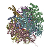
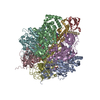
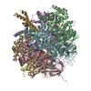
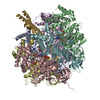
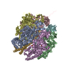
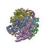
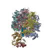
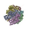
- PDB-2w6f: Low resolution structures of bovine mitochondrial F1-ATPase durin... -
+ Open data
- Basic information
| Entry | Database: PDB / ID: 2w6f | ||||||
|---|---|---|---|---|---|---|---|
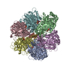
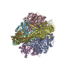
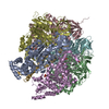
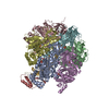
| Title | Low resolution structures of bovine mitochondrial F1-ATPase during controlled dehydration: Hydration State 2. | ||||||
 Components Components |
| ||||||
 Keywords Keywords | HYDROLASE / ATP PHOSPHORYLASE (H+ TRANSPORTING) / TRANSIT PEPTIDE / F1FO ATP SYNTHASE / ATP PHOSPHORYLASE / ATP SYNTHASE / ION TRANSPORT / MITOCHONDRION / ATP SYNTHESIS / UBL CONJUGATION / CF(1) / P-LOOP / NUCLEOTIDE-BINDING / HYDROGEN ION TRANSPORT / PYRROLIDONE CARBOXYLIC ACID / ATP-BINDING | ||||||
| Function / homology |  Function and homology information Function and homology informationFormation of ATP by chemiosmotic coupling / Cristae formation / Mitochondrial protein degradation / proton motive force-driven ATP synthesis / proton motive force-driven mitochondrial ATP synthesis / H+-transporting two-sector ATPase / proton-transporting ATP synthase complex / proton-transporting ATP synthase activity, rotational mechanism / ADP binding / mitochondrial inner membrane ...Formation of ATP by chemiosmotic coupling / Cristae formation / Mitochondrial protein degradation / proton motive force-driven ATP synthesis / proton motive force-driven mitochondrial ATP synthesis / H+-transporting two-sector ATPase / proton-transporting ATP synthase complex / proton-transporting ATP synthase activity, rotational mechanism / ADP binding / mitochondrial inner membrane / ATP hydrolysis activity / mitochondrion / ATP binding / plasma membrane Similarity search - Function | ||||||
| Biological species |  | ||||||
| Method |  X-RAY DIFFRACTION / X-RAY DIFFRACTION /  SYNCHROTRON / SYNCHROTRON /  MOLECULAR REPLACEMENT / Resolution: 6 Å MOLECULAR REPLACEMENT / Resolution: 6 Å | ||||||
 Authors Authors | Sanchez-Weatherby, J. / Felisaz, F. / Gobbo, A. / Huet, J. / Ravelli, R.B.G. / Bowler, M.W. / Cipriani, F. | ||||||
 Citation Citation |  Journal: Acta Crystallogr. D Biol. Crystallogr. / Year: 2009 Journal: Acta Crystallogr. D Biol. Crystallogr. / Year: 2009Title: Improving diffraction by humidity control: a novel device compatible with X-ray beamlines. Authors: Sanchez-Weatherby, J. / Bowler, M.W. / Huet, J. / Gobbo, A. / Felisaz, F. / Lavault, B. / Moya, R. / Kadlec, J. / Ravelli, R.B. / Cipriani, F. | ||||||
| History |
| ||||||
| Remark 700 | SHEET DETERMINATION METHOD: DSSP THE SHEETS PRESENTED AS "AA" IN EACH CHAIN ON SHEET RECORDS BELOW ... SHEET DETERMINATION METHOD: DSSP THE SHEETS PRESENTED AS "AA" IN EACH CHAIN ON SHEET RECORDS BELOW IS ACTUALLY AN 13-STRANDED BARREL THIS IS REPRESENTED BY A 14-STRANDED SHEET IN WHICH THE FIRST AND LAST STRANDS ARE IDENTICAL. THE SHEETS PRESENTED AS "BA", "CA" IN EACH CHAIN ON SHEET RECORDS BELOW IS ACTUALLY AN 11-STRANDED BARREL THIS IS REPRESENTED BY A 12-STRANDED SHEET IN WHICH THE FIRST AND LAST STRANDS ARE IDENTICAL. THE SHEET STRUCTURE OF THIS MOLECULE IS BIFURCATED. IN ORDER TO REPRESENT THIS FEATURE IN THE SHEET RECORDS BELOW, TWO SHEETS ARE DEFINED. |
- Structure visualization
| Structure viewer | Molecule:  Molmil Molmil Jmol/JSmol Jmol/JSmol |
|---|
- Downloads & links
- Download
| PDBx/mmCIF format |  2w6f.cif.gz 2w6f.cif.gz | 493.7 KB | Display |  PDBx/mmCIF format PDBx/mmCIF format |
|---|---|---|---|---|
| PDB format |  pdb2w6f.ent.gz pdb2w6f.ent.gz | 388.8 KB | Display |  PDB format PDB format |
| PDBx/mmJSON format |  2w6f.json.gz 2w6f.json.gz | Tree view |  PDBx/mmJSON format PDBx/mmJSON format | |
| Others |  Other downloads Other downloads |
-Validation report
| Arichive directory |  https://data.pdbj.org/pub/pdb/validation_reports/w6/2w6f https://data.pdbj.org/pub/pdb/validation_reports/w6/2w6f ftp://data.pdbj.org/pub/pdb/validation_reports/w6/2w6f ftp://data.pdbj.org/pub/pdb/validation_reports/w6/2w6f | HTTPS FTP |
|---|
-Related structure data
| Related structure data | 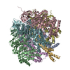 2w6eC 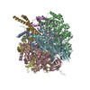 2w6gC 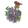 2w6hC  2w6iC  2w6jC 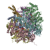 1bmfS S: Starting model for refinement C: citing same article ( |
|---|---|
| Similar structure data |
- Links
Links
- Assembly
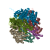
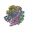
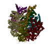
| Deposited unit | 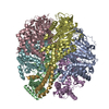
| ||||||||
|---|---|---|---|---|---|---|---|---|---|
| 1 |
| ||||||||
| Unit cell |
|
- Components
| #1: Protein | Mass: 59795.492 Da / Num. of mol.: 3 / Source method: isolated from a natural source / Source: (natural)  References: UniProt: P19483, H+-transporting two-sector ATPase #2: Protein | Mass: 56340.199 Da / Num. of mol.: 3 / Source method: isolated from a natural source / Source: (natural)  References: UniProt: P00829, H+-transporting two-sector ATPase #3: Protein | | Mass: 33119.035 Da / Num. of mol.: 1 / Source method: isolated from a natural source / Source: (natural)  References: UniProt: P05631, H+-transporting two-sector ATPase |
|---|
-Experimental details
-Experiment
| Experiment | Method:  X-RAY DIFFRACTION / Number of used crystals: 1 X-RAY DIFFRACTION / Number of used crystals: 1 |
|---|
- Sample preparation
| Crystal | Density Matthews: 3.34 Å3/Da / Density % sol: 62.83 % Description: THE DATA WERE COLLECTED AT ROOM TEMPERATURE, DURING CONTROLLED DEHYDRATION OF CRYSTALS,TO EVALUATE THE CHANGES THAT OCCUR IN CRYSTAL PACKING DURING DEHYDRATION. NO BIOLOGICAL ...Description: THE DATA WERE COLLECTED AT ROOM TEMPERATURE, DURING CONTROLLED DEHYDRATION OF CRYSTALS,TO EVALUATE THE CHANGES THAT OCCUR IN CRYSTAL PACKING DURING DEHYDRATION. NO BIOLOGICAL SIGNIFICANCE SHOULD BE ATTACHED TO THE COORDINATES. |
|---|---|
| Crystal grow | pH: 8.5 Details: 50 MM TRIS-HCL PH 8.2, 200 MM NACL, 20 MM MGSO4, 1 MM ADP, 1 MM ALCL3, 6 MM NAF 0.004% (W/V)PHENYLMETHYLSULFONYL FLUORIDE AND 12% (W/V) POLYETHYLENE GLYCOL 6000 |
-Data collection
| Diffraction | Mean temperature: 294 K |
|---|---|
| Diffraction source | Source:  SYNCHROTRON / Site: SYNCHROTRON / Site:  ESRF ESRF  / Beamline: ID14-2 / Wavelength: 0.933 / Beamline: ID14-2 / Wavelength: 0.933 |
| Detector | Type: ADSC CCD / Detector: CCD / Date: Oct 30, 2008 / Details: GE211 |
| Radiation | Monochromator: DIAMOND111 / Protocol: SINGLE WAVELENGTH / Monochromatic (M) / Laue (L): M / Scattering type: x-ray |
| Radiation wavelength | Wavelength: 0.933 Å / Relative weight: 1 |
| Reflection | Resolution: 6→101 Å / Num. obs: 10005 / % possible obs: 90.8 % / Observed criterion σ(I): 3 / Redundancy: 2.4 % / Rmerge(I) obs: 0.25 / Net I/σ(I): 7.9 |
| Reflection shell | Resolution: 6→6.32 Å / Redundancy: 2.3 % / Rmerge(I) obs: 0.82 / Mean I/σ(I) obs: 1.5 / % possible all: 91.7 |
- Processing
| Software |
| ||||||||||||||||||||
|---|---|---|---|---|---|---|---|---|---|---|---|---|---|---|---|---|---|---|---|---|---|
| Refinement | Method to determine structure:  MOLECULAR REPLACEMENT MOLECULAR REPLACEMENTStarting model: PDB ENTRY 1BMF Resolution: 6→30 Å / Cor.coef. Fo:Fc: 0.741 / Cor.coef. Fo:Fc free: 0.793 / SU B: 0.008 / SU ML: 0 / Cross valid method: THROUGHOUT / ESU R: 3.611 / ESU R Free: 3.591 / Stereochemistry target values: MAXIMUM LIKELIHOOD Details: HYDROGENS HAVE BEEN ADDED IN THE RIDING POSITIONS. THIS STRUCTURE WAS DETERMINED TO EVALUATE CHANGING CRYSTAL CONTACTS DURING CONTROLLED DEHYDRATION OF CRYSTALS. NO BIOLOGICAL SIGNIFICANCE ...Details: HYDROGENS HAVE BEEN ADDED IN THE RIDING POSITIONS. THIS STRUCTURE WAS DETERMINED TO EVALUATE CHANGING CRYSTAL CONTACTS DURING CONTROLLED DEHYDRATION OF CRYSTALS. NO BIOLOGICAL SIGNIFICANCE SHOULD BE ATTACHED TO THE COORDINATES.
| ||||||||||||||||||||
| Solvent computation | Ion probe radii: 0.8 Å / Shrinkage radii: 0.8 Å / VDW probe radii: 1.2 Å / Solvent model: MASK | ||||||||||||||||||||
| Displacement parameters | Biso mean: 20 Å2
| ||||||||||||||||||||
| Refinement step | Cycle: LAST / Resolution: 6→30 Å
| ||||||||||||||||||||
| LS refinement shell | Resolution: 6→6.15 Å / Total num. of bins used: 20 /
|
 Movie
Movie Controller


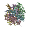
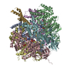

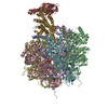








 PDBj
PDBj


