 Movie
Movie Controller

[English] 日本語
 Yorodumi
- PDB-1qo1: Molecular Architecture of the Rotary Motor in ATP Synthase from Y... -
+ Open data

- Basic information
| Entry | Database: PDB / ID: 1qo1 | ||||||
|---|---|---|---|---|---|---|---|
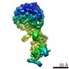
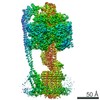
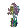
| Title | Molecular Architecture of the Rotary Motor in ATP Synthase from Yeast Mitochondria | ||||||
 Components Components |
| ||||||
 Keywords Keywords | ATP SYNTHASE / LOW RESOLUTION MODEL / C-ALPHA ONLY | ||||||
| Function / homology |  Function and homology information Function and homology information: / : / Formation of ATP by chemiosmotic coupling / Cristae formation / proton motive force-driven plasma membrane ATP synthesis / Mitochondrial protein degradation / proton motive force-driven ATP synthesis / proton-transporting two-sector ATPase complex, proton-transporting domain / proton motive force-driven mitochondrial ATP synthesis / proton-transporting ATPase activity, rotational mechanism ...: / : / Formation of ATP by chemiosmotic coupling / Cristae formation / proton motive force-driven plasma membrane ATP synthesis / Mitochondrial protein degradation / proton motive force-driven ATP synthesis / proton-transporting two-sector ATPase complex, proton-transporting domain / proton motive force-driven mitochondrial ATP synthesis / proton-transporting ATPase activity, rotational mechanism / membrane => GO:0016020 / H+-transporting two-sector ATPase / proton-transporting ATP synthase complex / proton-transporting ATP synthase activity, rotational mechanism / ADP binding / mitochondrial inner membrane / lipid binding / ATP hydrolysis activity / mitochondrion / ATP binding / metal ion binding / plasma membrane Similarity search - Function | ||||||
| Biological species |  | ||||||
| Method |  X-RAY DIFFRACTION / SYNCHROTRON / MOLECULAR REPLACEMENT / Resolution: 3.9 Å X-RAY DIFFRACTION / SYNCHROTRON / MOLECULAR REPLACEMENT / Resolution: 3.9 Å | ||||||
| Model type details | CA ATOMS ONLY, CHAIN A, B, C, D, E, F, G, J, K, L, M, N, O, P, Q, R, S, T | ||||||
 Authors Authors | Stock, D. / Leslie, A.G.W. / Walker, J.E. | ||||||
 Citation Citation | Journal: Science / Year: 1999 Title: Molecular Architecture of the Rotary Motor in ATP Synthase Authors: Stock, D. / Leslie, A.G.W. / Walker, J.E. #1: Journal: Biochemistry / Year: 1998Title: Solution Structure of the Transmembrane H -Transporting Subunit C of the F1F0 ATP Synthase Authors: Girvin, M.E. / Rastogi, V.K. / Abildgaard, F. / Markley, J.L. / Fillingame, R.H. #2: Journal: Structure / Year: 1997Title: Crystal Structure of the Epsilon Subunit of the Proton-Translocating ATP Synthase from Escherichia Coli Authors: Uhlin, U. / Cox, G.B. / Guss, J.M. #3: Journal: Nature / Year: 1994Title: Structure at 2.8-Angstrom Resolution of F1-ATPase from Bovine Heart Mitochondria Authors: Abrahams, J.P. / Leslie, A.G.W. / Lutter, R. / Walker, J.E. | ||||||
| History |
|
- Structure visualization
| Structure viewer | Molecule: MolmilJmol/JSmol |
|---|
- Downloads & links
-Download
| PDBx/mmCIF format | 1qo1.cif.gz | 134.3 KB | Display | PDBx/mmCIF format |
|---|---|---|---|---|
| PDB format | pdb1qo1.ent.gz | 87.1 KB | Display | PDB format |
| PDBx/mmJSON format | 1qo1.json.gz | Tree view | PDBx/mmJSON format | |
| Others |  Other downloads Other downloads |
-Validation report
| Arichive directory | https://data.pdbj.org/pub/pdb/validation_reports/qo/1qo1ftp://data.pdbj.org/pub/pdb/validation_reports/qo/1qo1 | HTTPS FTP |
|---|
-Related structure data
| Related structure data |  2xokC  1bmfS S: Starting model for refinement C: citing same article ( |
|---|---|
| Similar structure data |


-Links
 PDBj
PDBj


- Assembly
| Deposited unit | 
| ||||||||
|---|---|---|---|---|---|---|---|---|---|
| 1 |
| ||||||||
| Unit cell |
|
-Components
| #1: Protein | Mass: 55301.207 Da / Num. of mol.: 3 / Source method: isolated from a natural source Details: THE BOS TAURUS (BOVINE) ATP SYNTHASE ALPHA CHAIN HEART ISOFORM SEQUENCE (SWISSPROT P19483) AND MODEL (PDB ENTRY 1BMF) ARE GIVEN IN THE COORDINATES OF THIS ENTRY Source: (natural) #2: Protein | Mass: 51757.836 Da / Num. of mol.: 3 / Source method: isolated from a natural source Details: THE BOS TAURUS (BOVINE) ATP SYNTHASE BETA CHAIN SEQUENCE (SWISSPROT P00829) AND MODEL (PDB ENTRY 1BMF) ARE GIVEN IN THE COORDINATES OF THIS ENTRY Source: (natural) #3: Protein | | Mass: 30185.674 Da / Num. of mol.: 1 / Source method: isolated from a natural source Details: THE BOS TAURUS (BOVINE) ATP SYNTHASE GAMMA CHAIN SEQUENCE (SWISSPROT P05631) AND MODEL (PDB ENTRY 1BMF) ARE GIVEN IN THE COORDINATES OF THIS ENTRY Source: (natural) #4: Protein | | Mass: 14897.904 Da / Num. of mol.: 1 / Source method: isolated from a natural source Details: THE ESCHERICHIA COLI ATP SYNTHASE EPSILON CHAIN SEQUENCE (SWISSPROT P00832) AND MODEL (PDB ENTRY 1AQT) ARE GIVEN IN THE COORDINATES OF THIS ENTRY Source: (natural) References: UniProt: P00832, UniProt: P0A6E6*PLUS, EC: 3.6.1.34 #5: Protein | Mass: 8259.064 Da / Num. of mol.: 10 / Source method: isolated from a natural source Details: THE ESCHERICHIA COLI ATP SYNTHASE C CHAIN SEQUENCE (SWISSPROT P00844) AND MODEL (PDB ENTRY 1A91) ARE GIVEN IN THE COORDINATES OF THIS ENTRY Source: (natural) References: UniProt: P00844, UniProt: P68699*PLUS, EC: 3.6.1.34 Compound details | THE F-TYPE ATPASES HAVE 2 COMPONENTS, F1 - THE CATALYTIC CORE AND F0 - THE MEMBRANE PROTON CHANNEL. ...THE F-TYPE ATPASES HAVE 2 COMPONENTS | |
|---|
-Experimental details
-Experiment
| Experiment | Method: X-RAY DIFFRACTION / Number of used crystals: 1 |
|---|
- Sample preparation
| Crystal | Density Matthews: 3.66 Å3/Da / Density % sol: 66 % | ||||||||||||||||||||||||||||||||||||||||||||||||||||||
|---|---|---|---|---|---|---|---|---|---|---|---|---|---|---|---|---|---|---|---|---|---|---|---|---|---|---|---|---|---|---|---|---|---|---|---|---|---|---|---|---|---|---|---|---|---|---|---|---|---|---|---|---|---|---|---|
| Crystal grow | Method: microbatch / pH: 8 Details: 0.1 M TRIS/CL PH8.0, 12% PEG 6000, 150 MM NACL, 1 MM AMP-PNP, 40 MICROM ADP, 1 MM DTT, 0.02% NAN3, MIXED 1:1 WITH PROTEIN SOLUTION UNDER PARAFFIN OIL IN MICROBATCH PLATE., pH 8.00 | ||||||||||||||||||||||||||||||||||||||||||||||||||||||
| Crystal | *PLUS | ||||||||||||||||||||||||||||||||||||||||||||||||||||||
| Crystal grow | *PLUS Temperature: 4 ℃ / Method: batch method | ||||||||||||||||||||||||||||||||||||||||||||||||||||||
| Components of the solutions | *PLUS
|
-Data collection
| Diffraction | Mean temperature: 100 K |
|---|---|
| Diffraction source | Source: SYNCHROTRON / Site: ESRF  / Beamline: ID2 / Wavelength: 0.99 / Beamline: ID2 / Wavelength: 0.99 |
| Detector | Type: MARRESEARCH / Detector: IMAGE PLATE / Date: Feb 15, 1999 |
| Radiation | Protocol: SINGLE WAVELENGTH / Monochromatic (M) / Laue (L): M / Scattering type: x-ray |
| Radiation wavelength | Wavelength: 0.99 Å / Relative weight: 1 |
| Reflection | Resolution: 3.9→15 Å / Num. obs: 55593 / % possible obs: 93.3 % / Redundancy: 2.5 % / Rmerge(I) obs: 0.1 / Net I/σ(I): 4 |
| Reflection shell | Resolution: 3.9→4.11 Å / Redundancy: 2.5 % / Rmerge(I) obs: 0.37 / Mean I/σ(I) obs: 1.3 / % possible all: 95.5 |
| Reflection | *PLUS Rmerge(I) obs: 0.1 |
| Reflection shell | *PLUS % possible obs: 95.5 % / Num. unique obs: 8288 / Rmerge(I) obs: 0.372 |
- Processing
| Software |
| ||||||||||||
|---|---|---|---|---|---|---|---|---|---|---|---|---|---|
| Refinement | Method to determine structure: MOLECULAR REPLACEMENT Starting model: 1BMF Highest resolution: 3.9 Å Details: THE MODEL CONSISTS OF C ALPHA COORDINATES OF PDB ENTRIES 1BMF, 1AQT, AND 1A91, WHICH WERE FITTED INTO THE DENSITY AS RIGID BODIES. NO REFINEMENT WAS CARRIED OUT ON THIS MODEL. THE ELECTRON ...Details: THE MODEL CONSISTS OF C ALPHA COORDINATES OF PDB ENTRIES 1BMF, 1AQT, AND 1A91, WHICH WERE FITTED INTO THE DENSITY AS RIGID BODIES. NO REFINEMENT WAS CARRIED OUT ON THIS MODEL. THE ELECTRON DENSITY AT 3.9 ANGSTROM RESOLUTION DOES NOT PROVIDE THE INFORMATION REQUIRED TO DERIVE AN ATOMIC MODEL. THE COORDINATES ARE BASED ON COORDINATES OF MODELS OF SUBUNITS OR DOMAINS DERIVED FROM HIGHER RESOLUTION X-RAY EXPERIMENTS (1BMF, 1AQT) OR NMR- EXPERIMENTS (1A91). THESE MODELS WERE FITTED INTO THE DENSITY AS RIGID BODIES. THEREFORE THE RESIDUE NAMES OF THE C ALPHA ATOMS HAVE NO MEANING. | ||||||||||||
| Refinement step | Cycle: LAST / Highest resolution: 3.9 Å
| ||||||||||||
| Refinement | *PLUS Lowest resolution: 15 Å / Num. reflection obs: 55593 / Rfactor obs: 0.467 | ||||||||||||
| Solvent computation | *PLUS | ||||||||||||
| Displacement parameters | *PLUS | ||||||||||||
| LS refinement shell | *PLUS Rfactor obs: 0.506 |
