 Movie
Movie Controller

[English] 日本語
 Yorodumi
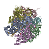
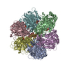
- PDB-1nbm: THE STRUCTURE OF BOVINE F1-ATPASE COVALENTLY INHIBITED WITH 4-CHL... -
+ Open data

- Basic information
| Entry | Database: PDB / ID: 1nbm | ||||||
|---|---|---|---|---|---|---|---|
| Title | THE STRUCTURE OF BOVINE F1-ATPASE COVALENTLY INHIBITED WITH 4-CHLORO-7-NITROBENZOFURAZAN | ||||||
 Components Components | (F1-ATPASE) x 4 | ||||||
 Keywords Keywords | ATP SYNTHASE / F1FO ATP SYNTHASE / F1-ATPASE / 4-CHLORO-7-NITROBENZOFURAZAN / INHIBITION | ||||||
| Function / homology |  Function and homology information Function and homology informationFormation of ATP by chemiosmotic coupling / Cristae formation / Mitochondrial protein degradation / proton motive force-driven ATP synthesis / proton motive force-driven mitochondrial ATP synthesis / H+-transporting two-sector ATPase / proton-transporting ATP synthase complex / proton-transporting ATP synthase activity, rotational mechanism / ADP binding / mitochondrial inner membrane ...Formation of ATP by chemiosmotic coupling / Cristae formation / Mitochondrial protein degradation / proton motive force-driven ATP synthesis / proton motive force-driven mitochondrial ATP synthesis / H+-transporting two-sector ATPase / proton-transporting ATP synthase complex / proton-transporting ATP synthase activity, rotational mechanism / ADP binding / mitochondrial inner membrane / ATP hydrolysis activity / mitochondrion / ATP binding / metal ion binding / plasma membrane Similarity search - Function | ||||||
| Biological species |  | ||||||
| Method |  X-RAY DIFFRACTION / SYNCHROTRON / MOLECULAR REPLACEMENT / Resolution: 3 Å X-RAY DIFFRACTION / SYNCHROTRON / MOLECULAR REPLACEMENT / Resolution: 3 Å | ||||||
 Authors Authors | Orriss, G.L. / Leslie, A.G.W. / Braig, K. / Walker, J.E. | ||||||
 Citation Citation | Journal: Structure / Year: 1998 Title: Bovine F1-ATPase covalently inhibited with 4-chloro-7-nitrobenzofurazan: the structure provides further support for a rotary catalytic mechanism. Authors: Orriss, G.L. / Leslie, A.G. / Braig, K. / Walker, J.E. #1: Journal: Nature / Year: 1994Title: Structure at 2.8 A Resolution of F1-ATPase from Bovine Heart Mitochondria Authors: Abrahams, J.P. / Leslie, A.G. / Lutter, R. / Walker, J.E. | ||||||
| History |
|
- Structure visualization
| Structure viewer | Molecule: MolmilJmol/JSmol |
|---|
- Downloads & links
-Download
| PDBx/mmCIF format | 1nbm.cif.gz | 573.9 KB | Display | PDBx/mmCIF format |
|---|---|---|---|---|
| PDB format | pdb1nbm.ent.gz | 465.3 KB | Display | PDB format |
| PDBx/mmJSON format | 1nbm.json.gz | Tree view | PDBx/mmJSON format | |
| Others |  Other downloads Other downloads |
-Validation report
| Arichive directory | https://data.pdbj.org/pub/pdb/validation_reports/nb/1nbmftp://data.pdbj.org/pub/pdb/validation_reports/nb/1nbm | HTTPS FTP |
|---|
-Related structure data
| Similar structure data |
|---|
-Links
 PDBj
PDBj


- Assembly
| Deposited unit | 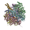
| ||||||||
|---|---|---|---|---|---|---|---|---|---|
| 1 |
| ||||||||
| Unit cell |
|
-Components
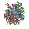
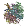
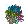
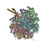
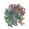
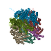
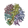
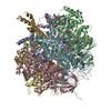
-Protein , 4 types, 7 molecules ABCDFEG
| #1: Protein | Mass: 55301.207 Da / Num. of mol.: 3 / Source method: isolated from a natural source / Source: (natural) #2: Protein | Mass: 51532.613 Da / Num. of mol.: 2 / Source method: isolated from a natural source / Source: (natural) #3: Protein | | Mass: 51667.738 Da / Num. of mol.: 1 / Source method: isolated from a natural source / Source: (natural) #4: Protein | | Mass: 30185.674 Da / Num. of mol.: 1 / Source method: isolated from a natural source / Source: (natural) |
|---|
-Non-polymers , 5 types, 175 molecules 


| #5: Chemical | ChemComp-MG /  Mass: 24.305 Da / Num. of mol.: 5 / Source method: obtained synthetically / Formula: Mg Mass: 24.305 Da / Num. of mol.: 5 / Source method: obtained synthetically / Formula: Mg#6: Chemical | ChemComp-ATP /  Mass: 507.181 Da / Num. of mol.: 4 / Source method: obtained synthetically / Formula: C10H16N5O13P3 / Comment: ATP, energy-carrying molecule*YM Mass: 507.181 Da / Num. of mol.: 4 / Source method: obtained synthetically / Formula: C10H16N5O13P3 / Comment: ATP, energy-carrying molecule*YM#7: Chemical | ChemComp-ADP / |  Mass: 427.201 Da / Num. of mol.: 1 / Source method: obtained synthetically / Formula: C10H15N5O10P2 / Comment: ADP, energy-carrying molecule*YM Mass: 427.201 Da / Num. of mol.: 1 / Source method: obtained synthetically / Formula: C10H15N5O10P2 / Comment: ADP, energy-carrying molecule*YM#8: Chemical | ChemComp-PO4 / |  Mass: 94.971 Da / Num. of mol.: 1 / Source method: obtained synthetically / Formula: PO4 Mass: 94.971 Da / Num. of mol.: 1 / Source method: obtained synthetically / Formula: PO4#9: Water | ChemComp-HOH / | Mass: 18.015 Da / Num. of mol.: 164 / Source method: isolated from a natural source / Formula: H2O |
|---|
-Details
| Compound details | THE F1-ATPASE MOLECULE HAS THREE COPIES OF THE NON-CATALYTIC ALPHA SUBUNIT AND THREE COPIES OF THE ...THE F1-ATPASE MOLECULE HAS THREE COPIES OF THE NON-CATALYTIC ALPHA SUBUNIT AND THREE COPIES OF THE CATALYTIC BETA SUBUNIT. IN THE PRIMARY REFERENCE, THE BETA SUBUNITS WERE LABELLED ACCORDING TO THE BOUND NUCLEOTIDE |
|---|
-Experimental details
-Experiment
| Experiment | Method: X-RAY DIFFRACTION / Number of used crystals: 1 |
|---|
- Sample preparation
| Crystal | Density Matthews: 2.8 Å3/Da / Density % sol: 56 % | ||||||||||||||||||||||||||||||||||||||||||||||||||||||||||||||||||||||||||||||||||||||||||||||||
|---|---|---|---|---|---|---|---|---|---|---|---|---|---|---|---|---|---|---|---|---|---|---|---|---|---|---|---|---|---|---|---|---|---|---|---|---|---|---|---|---|---|---|---|---|---|---|---|---|---|---|---|---|---|---|---|---|---|---|---|---|---|---|---|---|---|---|---|---|---|---|---|---|---|---|---|---|---|---|---|---|---|---|---|---|---|---|---|---|---|---|---|---|---|---|---|---|---|
| Crystal grow | pH: 7.5 Details: 50MM TRIS-HCL, PH7.5, 200MM SODIUM CHLORIDE, 20MM MAGNESIUM SULPHATE, 1MM EDTA, 0.002% (W/V) PHENYL METHYLSULPHONYL FLUORIDE, 0.02%(W/V) SODIUM AZIDE, 10.5% (W/V) PEG MME 5000, 250UM AMP-PNP AND 5UM ADP. | ||||||||||||||||||||||||||||||||||||||||||||||||||||||||||||||||||||||||||||||||||||||||||||||||
| Crystal | *PLUS | ||||||||||||||||||||||||||||||||||||||||||||||||||||||||||||||||||||||||||||||||||||||||||||||||
| Crystal grow | *PLUS Temperature: 23 ℃ / pH: 7.2 / Method: microdialysis | ||||||||||||||||||||||||||||||||||||||||||||||||||||||||||||||||||||||||||||||||||||||||||||||||
| Components of the solutions | *PLUS
|
-Data collection
| Diffraction | Mean temperature: 100 K |
|---|---|
| Diffraction source | Source: SYNCHROTRON / Site: SRS  / Beamline: PX9.6 / Wavelength: 0.87 / Beamline: PX9.6 / Wavelength: 0.87 |
| Detector | Type: MARRESEARCH / Detector: IMAGE PLATE / Date: May 12, 1997 |
| Radiation | Monochromatic (M) / Laue (L): M / Scattering type: x-ray |
| Radiation wavelength | Wavelength: 0.87 Å / Relative weight: 1 |
| Reflection | Resolution: 3→20 Å / Num. obs: 70240 / % possible obs: 97.2 % / Redundancy: 2.5 % / Biso Wilson estimate: 54.48 Å2 / Rmerge(I) obs: 0.088 / Net I/σ(I): 10.1 |
| Reflection shell | Resolution: 3→3.16 Å / Redundancy: 2.5 % / Rmerge(I) obs: 0.304 / Mean I/σ(I) obs: 3.4 / % possible all: 98 |
| Reflection shell | *PLUS % possible obs: 98 % |
- Processing
| Software |
| ||||||||||||||||||||||||||||||||||||||||||||||||||||||||||||
|---|---|---|---|---|---|---|---|---|---|---|---|---|---|---|---|---|---|---|---|---|---|---|---|---|---|---|---|---|---|---|---|---|---|---|---|---|---|---|---|---|---|---|---|---|---|---|---|---|---|---|---|---|---|---|---|---|---|---|---|---|---|
| Refinement | Method to determine structure: MOLECULAR REPLACEMENT Starting model: ALUMINUM FLUORIDE INHIBITED FORM OF BOVINE MITOCHONDRIAL F1-ATPASE Resolution: 3→6 Å / Isotropic thermal model: INDIVIDUAL / Cross valid method: FREE-R / σ(F): 2
| ||||||||||||||||||||||||||||||||||||||||||||||||||||||||||||
| Displacement parameters | Biso mean: 52.67 Å2 | ||||||||||||||||||||||||||||||||||||||||||||||||||||||||||||
| Refinement step | Cycle: LAST / Resolution: 3→6 Å
| ||||||||||||||||||||||||||||||||||||||||||||||||||||||||||||
| Refine LS restraints |
| ||||||||||||||||||||||||||||||||||||||||||||||||||||||||||||
| LS refinement shell | Resolution: 3→3.12 Å / Total num. of bins used: 8
| ||||||||||||||||||||||||||||||||||||||||||||||||||||||||||||
| Xplor file |
| ||||||||||||||||||||||||||||||||||||||||||||||||||||||||||||
| Software | *PLUS Name: X-PLOR / Version: 3.1 / Classification: refinement | ||||||||||||||||||||||||||||||||||||||||||||||||||||||||||||
| Refine LS restraints | *PLUS
|
