 Movie
Movie Controller

[English] 日本語
 Yorodumi
- PDB-1jnv: The Conformation of the Epsilon and Gamma Subunits within the E. ... -
+ Open data

- Basic information
| Entry | Database: PDB / ID: 1jnv | ||||||
|---|---|---|---|---|---|---|---|
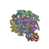
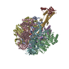
| Title | The Conformation of the Epsilon and Gamma Subunits within the E. coli F1 ATPase | ||||||
 Components Components |
| ||||||
 Keywords Keywords | HYDROLASE / F1 ATPase / ATP synthase / bioenergetics | ||||||
| Biological species |  | ||||||
| Method |  X-RAY DIFFRACTION / SYNCHROTRON / MOLECULAR REPLACEMENT / Resolution: 4.4 Å X-RAY DIFFRACTION / SYNCHROTRON / MOLECULAR REPLACEMENT / Resolution: 4.4 Å | ||||||
 Authors Authors | Hausrath, A.C. / Capaldi, R.A. / Matthews, B.W. | ||||||
 Citation Citation | Journal: J.Biol.Chem. / Year: 2001 Title: The conformation of the epsilon- and gamma-subunits within the Escherichia coli F(1) ATPase. Authors: Hausrath, A.C. / Capaldi, R.A. / Matthews, B.W. | ||||||
| History |
|
- Structure visualization
| Structure viewer | Molecule:  MolmilJmol/JSmol MolmilJmol/JSmol |
|---|
- Downloads & links
-Download
| PDBx/mmCIF format | 1jnv.cif.gz | 275.9 KB | Display | PDBx/mmCIF format |
|---|---|---|---|---|
| PDB format | pdb1jnv.ent.gz | 219.1 KB | Display | PDB format |
| PDBx/mmJSON format | 1jnv.json.gz | Tree view | PDBx/mmJSON format | |
| Others |  Other downloads Other downloads |
-Validation report
| Arichive directory | https://data.pdbj.org/pub/pdb/validation_reports/jn/1jnvftp://data.pdbj.org/pub/pdb/validation_reports/jn/1jnv | HTTPS FTP |
|---|
-Related structure data


-Links
 PDBj
PDBj
- Assembly
| Deposited unit | 
| ||||||||
|---|---|---|---|---|---|---|---|---|---|
| 1 |
| ||||||||
| Unit cell |
|
-Components
| #1: Protein | Mass: 41889.645 Da / Num. of mol.: 3 Source method: isolated from a genetically manipulated source Source: (gene. exp.) #2: Protein | Mass: 39762.000 Da / Num. of mol.: 3 Source method: isolated from a genetically manipulated source Source: (gene. exp.) #3: Protein | | Mass: 11507.176 Da / Num. of mol.: 1 Source method: isolated from a genetically manipulated source Source: (gene. exp.) #4: Protein | | Mass: 24442.998 Da / Num. of mol.: 1 Source method: isolated from a genetically manipulated source Source: (gene. exp.) Sequence details | THIS COMPOSITE MODEL WAS CONSTRUCTED BY PLACING COORDINATES FROM PDB ENTRIES 1BMF AND 1FSO INTO A ...THIS COMPOSITE MODEL WAS CONSTRUCTE | |
|---|
-Experimental details
-Experiment
| Experiment | Method: X-RAY DIFFRACTION / Number of used crystals: 1 |
|---|
- Sample preparation
| Crystal | Density Matthews: 2.9 Å3/Da / Density % sol: 60 % | ||||||||||||||||||||||||||||||||||||||||||||||||||||||||||||||||||||||||||||||
|---|---|---|---|---|---|---|---|---|---|---|---|---|---|---|---|---|---|---|---|---|---|---|---|---|---|---|---|---|---|---|---|---|---|---|---|---|---|---|---|---|---|---|---|---|---|---|---|---|---|---|---|---|---|---|---|---|---|---|---|---|---|---|---|---|---|---|---|---|---|---|---|---|---|---|---|---|---|---|---|
| Crystal grow | Temperature: 298 K / Method: vapor diffusion, hanging drop / pH: 7.2 Details: TRIS, PEG 8000, GLYCEROL, NACL, MGSO4, LISO4, NAN3, EDTA, AMP-PNP, ATP, pH 7.2, VAPOR DIFFUSION, HANGING DROP, temperature 298K | ||||||||||||||||||||||||||||||||||||||||||||||||||||||||||||||||||||||||||||||
| Crystal grow | *PLUS Temperature: 25 ℃Details: Hausrath, A.C., (1999) Proc.Natl.Acad.Sci.U.S.J., 96, 13697. | ||||||||||||||||||||||||||||||||||||||||||||||||||||||||||||||||||||||||||||||
| Components of the solutions | *PLUS
|
-Data collection
| Diffraction | Mean temperature: 100 K |
|---|---|
| Diffraction source | Source: SYNCHROTRON / Site: SSRL  / Beamline: BL9-1 / Wavelength: 0.98 Å / Beamline: BL9-1 / Wavelength: 0.98 Å |
| Detector | Type: MARRESEARCH / Detector: IMAGE PLATE |
| Radiation | Protocol: SINGLE WAVELENGTH / Monochromatic (M) / Laue (L): M / Scattering type: x-ray |
| Radiation wavelength | Wavelength: 0.98 Å / Relative weight: 1 |
| Reflection | Resolution: 4.4→25 Å / Num. all: 27196 / Num. obs: 17529 / % possible obs: 64 % / Redundancy: 5.1 % / Rmerge(I) obs: 0.122 / Net I/σ(I): 3.7 |
| Reflection shell | Resolution: 4.4→4.44 Å / Redundancy: 5.5 % / Rmerge(I) obs: 0.358 / Mean I/σ(I) obs: 2.1 / % possible all: 53.6 |
- Processing
| Software |
| ||||||||||||||||||||
|---|---|---|---|---|---|---|---|---|---|---|---|---|---|---|---|---|---|---|---|---|---|
| Refinement | Method to determine structure: MOLECULAR REPLACEMENT Starting model: PDB Entry 1bmf,1fs0 Resolution: 4.4→25 Å / σ(F): 0 / σ(I): 0 / Stereochemistry target values: TNT PROTGEO 1.0
| ||||||||||||||||||||
| Refinement step | Cycle: LAST / Resolution: 4.4→25 Å
| ||||||||||||||||||||
| Software | *PLUS Name: TNT / Classification: refinement | ||||||||||||||||||||
| Refinement | *PLUS Highest resolution: 4.4 Å / σ(F): 0 / Rfactor all: 0.416 | ||||||||||||||||||||
| Solvent computation | *PLUS | ||||||||||||||||||||
| Displacement parameters | *PLUS | ||||||||||||||||||||
| Refine LS restraints | *PLUS
|
