 Movie
Movie Controller


+ Open data

- Basic information
| Entry | Database: PDB / ID: 1gmg | ||||||
|---|---|---|---|---|---|---|---|
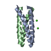
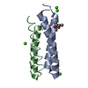
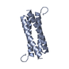
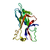
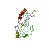
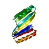
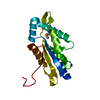
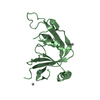
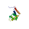
| Title | ALANINE 31 PROLINE MUTANT OF ROP PROTEIN, MONOCLINIC FORM | ||||||
 Components Components | REGULATORY PROTEIN ROP | ||||||
 Keywords Keywords | TRANSCRIPTION REGULATION | ||||||
| Function / homology | Helix Hairpins - #230 / Regulatory protein Rop / Rop-like superfamily / Rop protein / Helix Hairpins / Orthogonal Bundle / Mainly Alpha / identical protein binding / Regulatory protein rop Function and homology information Function and homology information | ||||||
| Biological species |  | ||||||
| Method |  X-RAY DIFFRACTION / MOLECULAR REPLACEMENT / Resolution: 1.9 Å X-RAY DIFFRACTION / MOLECULAR REPLACEMENT / Resolution: 1.9 Å | ||||||
 Authors Authors | Glykos, N.M. / Kokkinidis, M. | ||||||
 Citation Citation | Journal: Acta Crystallogr.,Sect.D / Year: 2003 Title: Structure Determination of a Small Protein Through a 23-Dimensional Molecular-Replacement Search. Authors: Glykos, N.M. / Kokkinidis, M. #1: Journal: Structure / Year: 1999Title: Protein Plasticity to the Extreme: Changing the Topology of a 4-Alpha-Helical Bundle with a Single Amino-Acid Substitution Authors: Glykos, N.M. / Cesareni, G. / Kokkinidis, M. #2: Journal: Acta Crystallogr.,Sect.D / Year: 1999 Title: Meaningful Refinement of Poly-Alanine Models Using Rigid-Body Simulated Annealing : Application to the Structure Determination of the A31P Rop Mutant Authors: Glykos, N.M. / Kokkinidis, M. | ||||||
| History |
|
- Structure visualization
| Structure viewer | Molecule: MolmilJmol/JSmol |
|---|
- Downloads & links
-Download
| PDBx/mmCIF format | 1gmg.cif.gz | 36.1 KB | Display | PDBx/mmCIF format |
|---|---|---|---|---|
| PDB format | pdb1gmg.ent.gz | 24.6 KB | Display | PDB format |
| PDBx/mmJSON format | 1gmg.json.gz | Tree view | PDBx/mmJSON format | |
| Others |  Other downloads Other downloads |
-Validation report
| Arichive directory | https://data.pdbj.org/pub/pdb/validation_reports/gm/1gmgftp://data.pdbj.org/pub/pdb/validation_reports/gm/1gmg | HTTPS FTP |
|---|
-Related structure data
| Related structure data | 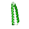 1rpoS S: Starting model for refinement |
|---|---|
| Similar structure data |


-Links
 PDBj
PDBj- Assembly
| Deposited unit | 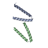
| ||||||||||||
|---|---|---|---|---|---|---|---|---|---|---|---|---|---|
| 1 | 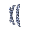
| ||||||||||||
| 2 | 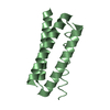
| ||||||||||||
| Unit cell |
| ||||||||||||
| Components on special symmetry positions |
| ||||||||||||
| Noncrystallographic symmetry (NCS) | NCS oper: (Code: given Matrix: (-0.99579, 0.02097, 0.0892), Vector: Details | THE CRYSTALLOGRAPHIC ASYMMETRIC UNIT CONTAINS TWOCRYSTALLOGRAPHICALLY INDEPENDENT MONOMERS (HALF BUNDLES).THE TWO COMPLETE 4-ALPHA-HELICAL BUNDLES ARE FORMED THROUGHTHE APPLICATION OF CRYSTALLOGRAPHIC SYMMETRY OPERATORSCORRESPONDING TO THE TWO-FOLD AXES (PARALLEL TO Y) ATX=0.00, Z= 0.00 AND AT X=0.50, Z=0.50.THE CHAIN IN THE COMPLEX ARE 1268.5 ANGSTROM**2AND 1158. 3 ANGSTROM**2 | |
-Components
| #1: Protein | Mass: 7263.076 Da / Num. of mol.: 2 / Mutation: YES Source method: isolated from a genetically manipulated source Source: (gene. exp.) #2: Water | ChemComp-HOH / |  Mass: 18.015 Da / Num. of mol.: 92 / Source method: isolated from a natural source / Formula: H2O Mass: 18.015 Da / Num. of mol.: 92 / Source method: isolated from a natural source / Formula: H2OCompound details | CHAIN A, B ENGINEERED | Has protein modification | Y | |
|---|
-Experimental details
-Experiment
| Experiment | Method: X-RAY DIFFRACTION / Number of used crystals: 1 |
|---|
- Sample preparation
| Crystal | Density Matthews: 2.13 Å3/Da / Density % sol: 41.8 % Description: THE STRUCTURE WAS DETERMINED THROUGH A 23-DIMENSIONAL MOLECULAR REPLACEMENT SEARCH PERFORMED WITH THE PROGRAM QUEEN OF SPADES AND USING AS A SEARCH MODEL ONE POLY-ALANINE HELIX ...Description: THE STRUCTURE WAS DETERMINED THROUGH A 23-DIMENSIONAL MOLECULAR REPLACEMENT SEARCH PERFORMED WITH THE PROGRAM QUEEN OF SPADES AND USING AS A SEARCH MODEL ONE POLY-ALANINE HELIX ACCOUNTING FOR 13% OF THE TOTAL NUMBER OF ATOMS. |
|---|---|
| Crystal grow | pH: 4.8 Details: 37% (V/V) ETHANOL, 450 MM NACL, 50 MM CITRATE BUFFER PH4.8, 1MM EDTA, USING THE DIALYSIS METHOD., pH 4.80 |
| Crystal grow | *PLUS Method: other / Details: Glykos, N.M., (1999) Acta Cryst., D55, 1301. |
-Data collection
| Diffraction | Mean temperature: 293 K |
|---|---|
| Diffraction source | Source: ROTATING ANODE / Type: RIGAKU RU300 / Wavelength: 1.5418 |
| Detector | Type: MARRESEARCH / Detector: IMAGE PLATE / Date: Nov 15, 1999 / Details: YALE MIRRORS |
| Radiation | Monochromator: YALE MIRRORS / Protocol: SINGLE WAVELENGTH / Monochromatic (M) / Laue (L): M / Scattering type: x-ray |
| Radiation wavelength | Wavelength: 1.5418 Å / Relative weight: 1 |
| Reflection | Resolution: 1.9→36 Å / Num. obs: 8771 / % possible obs: 96.4 % / Redundancy: 8.2 % / Biso Wilson estimate: 39.6 Å2 / Rmerge(I) obs: 0.075 / Net I/σ(I): 12.4 |
| Reflection shell | Resolution: 1.9→1.97 Å / Redundancy: 4.1 % / Rmerge(I) obs: 0.291 / Mean I/σ(I) obs: 4.1 / % possible all: 91.6 |
| Reflection | *PLUS Highest resolution: 1.9 Å / Lowest resolution: 36 Å |
| Reflection shell | *PLUS % possible obs: 91.6 % |
- Processing
| Software |
| ||||||||||||||||||||||||||||||||||||||||||||||||||||||||||||
|---|---|---|---|---|---|---|---|---|---|---|---|---|---|---|---|---|---|---|---|---|---|---|---|---|---|---|---|---|---|---|---|---|---|---|---|---|---|---|---|---|---|---|---|---|---|---|---|---|---|---|---|---|---|---|---|---|---|---|---|---|---|
| Refinement | Method to determine structure: MOLECULAR REPLACEMENT Starting model: POLY-ALANINE MODEL OF ONE HELIX (RESIDUES 4-29) FROM PDB ENTRY 1RPO Resolution: 1.9→36 Å / Isotropic thermal model: RESTRAINED / Cross valid method: THROUGHOUT / σ(F): 0 Details: RESOLUTION-DEPENDENT TWO-LINE WEIGHTING SCHEME (DAVID SMITH, G. (1997), ACTA CRYST. D53, PP.41-48).
| ||||||||||||||||||||||||||||||||||||||||||||||||||||||||||||
| Displacement parameters | Biso mean: 41.16 Å2
| ||||||||||||||||||||||||||||||||||||||||||||||||||||||||||||
| Refine analyze | Luzzati coordinate error obs: 0.19 Å / Luzzati d res low obs: 36 Å | ||||||||||||||||||||||||||||||||||||||||||||||||||||||||||||
| Refinement step | Cycle: LAST / Resolution: 1.9→36 Å
| ||||||||||||||||||||||||||||||||||||||||||||||||||||||||||||
| Refine LS restraints |
| ||||||||||||||||||||||||||||||||||||||||||||||||||||||||||||
| LS refinement shell | Resolution: 1.9→1.99 Å / Total num. of bins used: 8
| ||||||||||||||||||||||||||||||||||||||||||||||||||||||||||||
| Xplor file |
| ||||||||||||||||||||||||||||||||||||||||||||||||||||||||||||
| Refinement | *PLUS Highest resolution: 1.9 Å / Lowest resolution: 36 Å | ||||||||||||||||||||||||||||||||||||||||||||||||||||||||||||
| Solvent computation | *PLUS | ||||||||||||||||||||||||||||||||||||||||||||||||||||||||||||
| Displacement parameters | *PLUS |
