[English] 日本語
Yorodumi
- PDB-1b6q: ALANINE 31 PROLINE MUTANT OF ROP PROTEIN -

+
Open data


ID or keywords:

Loading...

-
Basic information

Entry
Database: PDB / ID: 1b6q
TitleALANINE 31 PROLINE MUTANT OF ROP PROTEIN
ComponentsROP
KeywordsTRANSCRIPTION REGULATION
Function / homologyHelix Hairpins - #230 / Regulatory protein Rop / Rop-like superfamily / Rop protein / Helix Hairpins / Orthogonal Bundle / Mainly Alpha / identical protein binding / Regulatory protein rop
Function and homology information
Biological speciesEscherichia coli (E. coli)
MethodX-RAY DIFFRACTION / MOLECULAR REPLACEMENT / Resolution: 1.8 Å
AuthorsGlykos, N. / Cesareni, G. / Kokkinidis, M.
Citation
Journal: Structure Fold.Des. / Year: 1999
Title: Protein plasticity to the extreme: changing the topology of a 4-alpha-helical bundle with a single amino acid substitution.
Authors: Glykos, N.M. / Cesareni, G. / Kokkinidis, M.
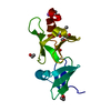
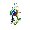
#1: Journal: Acta Crystallogr.,Sect.D / Year: 1999
Title: Meaningful Refinement of Poly-Alanine Models Using Rigid-Body Simulated Annealing : Application to the Structure Determination of the A31P Rop Mutant
History
DepositionJan 16, 1999Processing site: BNL
Revision 1.0Jul 9, 1999Provider: repository / Type: Initial release
Revision 1.1Mar 24, 2008Group: Version format compliance
Revision 1.2Jul 13, 2011Group: Derived calculations / Version format compliance
Revision 1.3Nov 16, 2011Group: Atomic model
Revision 1.4Apr 4, 2018Group: Data collection / Other / Category: diffrn_source / pdbx_database_status
Item: _diffrn_source.type / _pdbx_database_status.process_site
Revision 1.5Apr 18, 2018Group: Data collection / Category: diffrn_detector / Item: _diffrn_detector.detector
Revision 1.6Aug 2, 2023Group: Database references / Refinement description
Category: database_2 / pdbx_initial_refinement_model / struct_ref_seq_dif
Item: _database_2.pdbx_DOI / _database_2.pdbx_database_accession / _struct_ref_seq_dif.details
Revision 1.7May 22, 2024Group: Data collection / Category: chem_comp_atom / chem_comp_bond

-
Structure visualization

Structure viewerMolecule:
MolmilJmol/JSmol

Downloads & links

-
Assembly

Deposited unit
A: ROP


Theoretical massNumber of molelcules
Total (without water)7,2631
Polymers7,2631
Non-polymers00
Water99155
1
A: ROP

A: ROP


Theoretical massNumber of molelcules
Total (without water)14,5262
Polymers14,5262
Non-polymers00
Water362
TypeNameSymmetry operationNumber
identity operation1_555x,y,z1
crystal symmetry operation3_555-x,y,-z+1/21
Buried area3070 Å2
ΔGint-38 kcal/mol
Surface area7350 Å2
MethodPISA, PQS
Unit cell
Length a, b, c (Å)30.400, 42.100, 81.400
Angle α, β, γ (deg.)90.00, 90.00, 90.00
Int Tables number20
Space group name H-MC2221
Components on special symmetry positions
IDModelComponents
11A-103-

HOH

21A-143-

HOH

-
Components

#1: Protein ROP / ROM


Mass: 7263.076 Da / Num. of mol.: 1 / Mutation: A31P
Source method: isolated from a genetically manipulated source
Source: (gene. exp.) Escherichia coli (E. coli) / Cellular location: CYTOPLASM / Gene: ROP / Plasmid: PEX43 / Production host: Escherichia coli (E. coli) / Strain (production host): 71/72 (71/18 PLUS PCI857) / References: UniProt: P03051
#2: Water ChemComp-HOH / water / Water


Mass: 18.015 Da / Num. of mol.: 55 / Source method: isolated from a natural source / Formula: H2O

-
Experimental details

-
Experiment

ExperimentMethod: X-RAY DIFFRACTION / Number of used crystals: 1

-
Sample preparation

CrystalDensity Matthews: 1.8 Å3/Da / Density % sol: 35 %
Crystal growpH: 4.3 / Details: pH 4.3
Crystal grow
*PLUS
Method: vapor diffusion, hanging drop / PH range low: 5.4 / PH range high: 5
Components of the solutions
*PLUS
IDConc.Common nameCrystal-IDSol-ID
115 mg/mlprotein1drop
2100 mMsodium acetate1drop
3100 mMammonium sulfate1drop
41.6 Mammonium sulfate1reservoir

-
Data collection

DiffractionMean temperature: 293 K
Diffraction sourceSource: SEALED TUBE / Type: OTHER / Wavelength: 1.5418
DetectorType: ENRAF-NONIUS FAST / Detector: DIFFRACTOMETER / Details: MONOCHROMATOR
RadiationMonochromator: GRAPHITE(002) / Monochromatic (M) / Laue (L): M / Scattering type: x-ray
Radiation wavelengthWavelength: 1.5418 Å / Relative weight: 1
ReflectionResolution: 1.8→40.825 Å / Num. obs: 5103 / % possible obs: 99.9 % / Redundancy: 1 % / Biso Wilson estimate: 28.9 Å2 / Net I/σ(I): 12.4
Reflection shellResolution: 1.8→1.86 Å / Redundancy: 1 % / Mean I/σ(I) obs: 2.48 / % possible all: 99.6
Reflection shell
*PLUS
% possible obs: 99.6 %

-
Processing

Software
NameClassification
X-PLORmodel building
X-PLORrefinement
CAD4data reduction
CAD4data scaling
X-PLORphasing
RefinementMethod to determine structure: MOLECULAR REPLACEMENT
Starting model: POLY-ALANINE MODEL FROM PDB ENTRY 1ROP
Resolution: 1.8→40.825 Å / Isotropic thermal model: RESTRAINED / Cross valid method: THROUGHOUT / σ(F): 0
Details: RESOLUTION-DEPENDENT TWO-LINE WEIGHTING SCHEME AS SUGGESTED BY DAVID SMITH, PROCEEDINGS OF THE CCP4 STUDY WEEKEND, 1996.
RfactorNum. reflection% reflectionSelection details
Rfree0.24 286 5 %RANDOM
Rwork0.189 ---
obs0.189 5103 99.9 %-
Displacement parametersBiso mean: 26 Å2
Baniso -1Baniso -2Baniso -3
1-6.2422 Å20 Å20 Å2
2---1.7673 Å20 Å2
3----4.4617 Å2
Refine analyzeLuzzati coordinate error obs: 0.23 Å / Luzzati d res low obs: 40.8 Å
Refinement stepCycle: LAST / Resolution: 1.8→40.825 Å
ProteinNucleic acidLigandSolventTotal
Num. atoms440 0 0 55 495
Refine LS restraints
Refine-IDTypeDev ideal
X-RAY DIFFRACTIONx_bond_d0.007
X-RAY DIFFRACTIONx_bond_d_na
X-RAY DIFFRACTIONx_bond_d_prot
X-RAY DIFFRACTIONx_angle_d
X-RAY DIFFRACTIONx_angle_d_na
X-RAY DIFFRACTIONx_angle_d_prot
X-RAY DIFFRACTIONx_angle_deg0.909
X-RAY DIFFRACTIONx_angle_deg_na
X-RAY DIFFRACTIONx_angle_deg_prot
X-RAY DIFFRACTIONx_dihedral_angle_d
X-RAY DIFFRACTIONx_dihedral_angle_d_na
X-RAY DIFFRACTIONx_dihedral_angle_d_prot
X-RAY DIFFRACTIONx_improper_angle_d
X-RAY DIFFRACTIONx_improper_angle_d_na
X-RAY DIFFRACTIONx_improper_angle_d_prot
X-RAY DIFFRACTIONx_mcbond_it1.53
X-RAY DIFFRACTIONx_mcangle_it
X-RAY DIFFRACTIONx_scbond_it2.73
X-RAY DIFFRACTIONx_scangle_it
LS refinement shellResolution: 1.8→1.88 Å / Total num. of bins used: 8
RfactorNum. reflection% reflection
Rfree0.4 31 5 %
Rwork0.332 588 -
obs--99.9 %
Xplor file
Refine-IDSerial noParam file
X-RAY DIFFRACTION1TOPHCSDX.PRO
X-RAY DIFFRACTION2TOPH19.SOL
X-RAY DIFFRACTION3PROTEIN_REP.PARAM
Software
*PLUS
Name: X-PLOR / Version: 3.851 / Classification: refinement
Refinement
*PLUS
Rfactor obs: 0.188
Solvent computation
*PLUS
Displacement parameters
*PLUS

+
About Yorodumi

-
News

-
Feb 9, 2022. New format data for meta-information of EMDB entries

New format data for meta-information of EMDB entries

  • Version 3 of the EMDB header file is now the official format.
  • The previous official version 1.9 will be removed from the archive.

Related info.:EMDB header

External links:wwPDB to switch to version 3 of the EMDB data model

-
Aug 12, 2020. Covid-19 info

Covid-19 info

URL: https://pdbj.org/emnavi/covid19.php

New page: Covid-19 featured information page in EM Navigator.

Related info.:Covid-19 info / Mar 5, 2020. Novel coronavirus structure data

+
Mar 5, 2020. Novel coronavirus structure data

Novel coronavirus structure data

Related info.:Yorodumi Speices / Aug 12, 2020. Covid-19 info

External links:COVID-19 featured content - PDBj / Molecule of the Month (242):Coronavirus Proteases

+
Jan 31, 2019. EMDB accession codes are about to change! (news from PDBe EMDB page)

EMDB accession codes are about to change! (news from PDBe EMDB page)

  • The allocation of 4 digits for EMDB accession codes will soon come to an end. Whilst these codes will remain in use, new EMDB accession codes will include an additional digit and will expand incrementally as the available range of codes is exhausted. The current 4-digit format prefixed with “EMD-” (i.e. EMD-XXXX) will advance to a 5-digit format (i.e. EMD-XXXXX), and so on. It is currently estimated that the 4-digit codes will be depleted around Spring 2019, at which point the 5-digit format will come into force.
  • The EM Navigator/Yorodumi systems omit the EMD- prefix.

Related info.:Q: What is EMD? / ID/Accession-code notation in Yorodumi/EM Navigator

External links:EMDB Accession Codes are Changing Soon! / Contact to PDBj

+
Jul 12, 2017. Major update of PDB

Major update of PDB

  • wwPDB released updated PDB data conforming to the new PDBx/mmCIF dictionary.
  • This is a major update changing the version number from 4 to 5, and with Remediation, in which all the entries are updated.
  • In this update, many items about electron microscopy experimental information are reorganized (e.g. em_software).
  • Now, EM Navigator and Yorodumi are based on the updated data.

External links:wwPDB Remediation / Enriched Model Files Conforming to OneDep Data Standards Now Available in the PDB FTP Archive

-
Yorodumi

Thousand views of thousand structures

  • Yorodumi is a browser for structure data from EMDB, PDB, SASBDB, etc.
  • This page is also the successor to EM Navigator detail page, and also detail information page/front-end page for Omokage search.
  • The word "yorodu" (or yorozu) is an old Japanese word meaning "ten thousand". "mi" (miru) is to see.

Related info.:EMDB / PDB / SASBDB / Comparison of 3 databanks / Yorodumi Search / Aug 31, 2016. New EM Navigator & Yorodumi / Yorodumi Papers / Jmol/JSmol / Function and homology information / Changes in new EM Navigator and Yorodumi

Read more