 Movie
Movie Controller

[English] 日本語
 Yorodumi
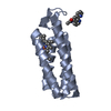
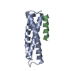
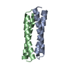
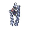
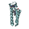
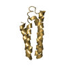
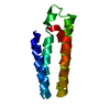
- PDB-1nkd: ATOMIC RESOLUTION (1.07 ANGSTROMS) STRUCTURE OF THE ROP MUTANT <2AA> -
+ Open data

- Basic information
| Entry | Database: PDB / ID: 1nkd | ||||||
|---|---|---|---|---|---|---|---|
| Title | ATOMIC RESOLUTION (1.07 ANGSTROMS) STRUCTURE OF THE ROP MUTANT <2AA> | ||||||
 Components Components | ROP | ||||||
 Keywords Keywords | TRANSCRIPTION REGULATION / ROP (COLE1 REPRESSOR OF PRIMER) / ATOMIC RESOLUTION STRUCTURE / 4-ALPHA-HELIX BUNDLE | ||||||
| Function / homology | Helix Hairpins - #230 / Regulatory protein Rop / Rop-like superfamily / Rop protein / Helix Hairpins / Orthogonal Bundle / Mainly Alpha / identical protein binding / Regulatory protein rop Function and homology information Function and homology information | ||||||
| Biological species |  | ||||||
| Method |  X-RAY DIFFRACTION / SYNCHROTRON / MOLECULAR REPLACEMENT / Resolution: 1.09 Å X-RAY DIFFRACTION / SYNCHROTRON / MOLECULAR REPLACEMENT / Resolution: 1.09 Å | ||||||
 Authors Authors | Vlassi, M. / Kokkinidis, M. | ||||||
 Citation Citation | Journal: Acta Crystallogr.,Sect.D / Year: 1998 Title: Structural parameters for proteins derived from the atomic resolution (1.09 A) structure of a designed variant of the ColE1 ROP protein. Authors: Vlassi, M. / Dauter, Z. / Wilson, K.S. / Kokkinidis, M. #1: Journal: Nat.Struct.Biol. / Year: 1994Title: Restored Heptad Pattern Continuity Does not Alter the Folding of a Four-Alpha-Helix Bundle Authors: Vlassi, M. / Steif, C. / Weber, P. / Tsernoglou, D. / Wilson, K.S. / Hinz, H.J. / Kokkinidis, M. #2: Journal: Proteins / Year: 1993Title: Correlation between Protein Stability and Crystal Properties of Designed Rop Variants Authors: Kokkinidis, M. / Vlassi, M. / Papanikolaou, Y. / Kotsifaki, D. / Kingswell, A. / Tsernoglou, D. / Hinz, H.J. #3: Journal: J.Mol.Biol. / Year: 1987Title: Structure of the Cole1 Rop Protein at 1.7 A Resolution Authors: Banner, D.W. / Kokkinidis, M. / Tsernoglou, D. | ||||||
| History |
|
- Structure visualization
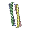
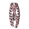
| Structure viewer | Molecule: MolmilJmol/JSmol |
|---|
- Downloads & links
-Download
| PDBx/mmCIF format | 1nkd.cif.gz | 37.3 KB | Display | PDBx/mmCIF format |
|---|---|---|---|---|
| PDB format | pdb1nkd.ent.gz | 26.2 KB | Display | PDB format |
| PDBx/mmJSON format | 1nkd.json.gz | Tree view | PDBx/mmJSON format | |
| Others |  Other downloads Other downloads |
-Validation report
| Arichive directory | https://data.pdbj.org/pub/pdb/validation_reports/nk/1nkdftp://data.pdbj.org/pub/pdb/validation_reports/nk/1nkd | HTTPS FTP |
|---|
-Related structure data
| Similar structure data |
|---|

-Links
 PDBj
PDBj- Assembly
| Deposited unit | 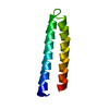
| ||||||||
|---|---|---|---|---|---|---|---|---|---|
| 1 | 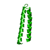
| ||||||||
| Unit cell |
| ||||||||
| Components on special symmetry positions |
|
-Components
| #1: Protein | Mass: 7379.194 Da / Num. of mol.: 1 / Mutation: INS(A-D31-A) Source method: isolated from a genetically manipulated source Source: (gene. exp.) |
|---|---|
| #2: Water | ChemComp-HOH /  Mass: 18.015 Da / Num. of mol.: 114 / Source method: isolated from a natural source / Formula: H2O Mass: 18.015 Da / Num. of mol.: 114 / Source method: isolated from a natural source / Formula: H2O |
-Experimental details
-Experiment
| Experiment | Method: X-RAY DIFFRACTION / Number of used crystals: 1 |
|---|
- Sample preparation
| Crystal | Density Matthews: 1.9 Å3/Da / Density % sol: 35 % | |||||||||||||||||||||||||
|---|---|---|---|---|---|---|---|---|---|---|---|---|---|---|---|---|---|---|---|---|---|---|---|---|---|---|
| Crystal grow | *PLUS Method: vapor diffusion, hanging drop / PH range low: 5.4 / PH range high: 5 | |||||||||||||||||||||||||
| Components of the solutions | *PLUS
|
-Data collection
| Diffraction source | Source: SYNCHROTRON / Site: EMBL/DESY, HAMBURG  / Beamline: X11 / Wavelength: 0.92 / Beamline: X11 / Wavelength: 0.92 |
|---|---|
| Radiation | Monochromatic (M) / Laue (L): M / Scattering type: x-ray |
| Radiation wavelength | Wavelength: 0.92 Å / Relative weight: 1 |
| Reflection | Resolution: 1.09→23.13 Å / Num. obs: 22361 / % possible obs: 98.2 % / Redundancy: 3.7 % / Rmerge(I) obs: 0.45 / Net I/σ(I): 18.9 |
| Reflection shell | Resolution: 1.09→1.19 Å / Redundancy: 2.5 % / Rmerge(I) obs: 0.197 / Mean I/σ(I) obs: 8.2 / % possible all: 97.5 |
| Reflection | *PLUS Biso Wilson estimate: 9.7 Å2 |
| Reflection shell | *PLUS % possible obs: 97.5 % |
- Processing
| Software |
| |||||||||||||||||||||||||||||||||
|---|---|---|---|---|---|---|---|---|---|---|---|---|---|---|---|---|---|---|---|---|---|---|---|---|---|---|---|---|---|---|---|---|---|---|
| Refinement | Method to determine structure: MOLECULAR REPLACEMENT / Resolution: 1.09→8 Å / Num. parameters: 5660 / Num. restraintsaints: 5406 / Cross valid method: FREE R / σ(F): 0 / Stereochemistry target values: ENGH & HUBER, 1991.
| |||||||||||||||||||||||||||||||||
| Refine analyze | Num. disordered residues: 10 / Occupancy sum hydrogen: 1 / Occupancy sum non hydrogen: 1 | |||||||||||||||||||||||||||||||||
| Refinement step | Cycle: LAST / Resolution: 1.09→8 Å
| |||||||||||||||||||||||||||||||||
| Refine LS restraints |
|
