 Movie
Movie Controller

[English] 日本語
 Yorodumi
- PDB-4do2: Crystal Structure of the Rop protein mutant D30P/A31G at resoluti... -
+ Open data

- Basic information
| Entry | Database: PDB / ID: 4do2 | ||||||
|---|---|---|---|---|---|---|---|
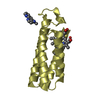
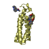
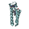
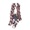
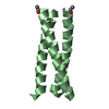
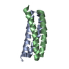
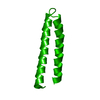
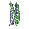
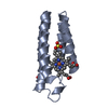
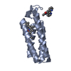
| Title | Crystal Structure of the Rop protein mutant D30P/A31G at resolution 1.4 resolution. | ||||||
 Components Components | Regulatory protein rop | ||||||
 Keywords Keywords | RNA BINDING PROTEIN / protein structure / protein folding / Rop protein / bacterial protein / mutation / 4-alpha-helical bundle / loop / ColE1 plasmid copy number | ||||||
| Function / homology | Helix Hairpins - #230 / Regulatory protein Rop / Rop-like superfamily / Rop protein / Helix Hairpins / Orthogonal Bundle / Mainly Alpha / identical protein binding / Regulatory protein rop Function and homology information Function and homology information | ||||||
| Biological species |  | ||||||
| Method |  X-RAY DIFFRACTION / SYNCHROTRON / MOLECULAR REPLACEMENT / Resolution: 1.401 Å X-RAY DIFFRACTION / SYNCHROTRON / MOLECULAR REPLACEMENT / Resolution: 1.401 Å | ||||||
 Authors Authors | Amprazi, M. / Kapetaniou, E.G. / Kokkinidis, M. | ||||||
 Citation Citation | Journal: Proc.Natl.Acad.Sci.USA / Year: 2014 Title: Structural plasticity of 4-alpha-helical bundles exemplified by the puzzle-like molecular assembly of the Rop protein. Authors: Amprazi, M. / Kotsifaki, D. / Providaki, M. / Kapetaniou, E.G. / Fellas, G. / Kyriazidis, I. / Perez, J. / Kokkinidis, M. #1: Journal: Acta Crystallogr.,Sect.F / Year: 2008 Title: Purification, crystallization and preliminary X-ray diffraction analysis of a variant of the ColE1 Rop protein. Authors: Ambrazi, M. / Fellas, G. / Kapetaniou, E.G. / Kotsifaki, D. / Providaki, M. / Kokkinidis, M. | ||||||
| History |
|
- Structure visualization
| Structure viewer | Molecule: MolmilJmol/JSmol |
|---|
- Downloads & links
-Download
| PDBx/mmCIF format | 4do2.cif.gz | 39.5 KB | Display | PDBx/mmCIF format |
|---|---|---|---|---|
| PDB format | pdb4do2.ent.gz | 27.3 KB | Display | PDB format |
| PDBx/mmJSON format | 4do2.json.gz | Tree view | PDBx/mmJSON format | |
| Others |  Other downloads Other downloads |
-Validation report
| Arichive directory | https://data.pdbj.org/pub/pdb/validation_reports/do/4do2ftp://data.pdbj.org/pub/pdb/validation_reports/do/4do2 | HTTPS FTP |
|---|
-Related structure data
| Related structure data |  1ropS S: Starting model for refinement |
|---|---|
| Similar structure data |
-Links
 PDBj
PDBj- Assembly
| Deposited unit | 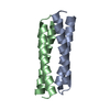
| ||||||||
|---|---|---|---|---|---|---|---|---|---|
| 1 |
| ||||||||
| Unit cell |
|
-Components
| #1: Protein | Mass: 8163.035 Da / Num. of mol.: 2 / Mutation: D30P, A31G Source method: isolated from a genetically manipulated source Source: (gene. exp.) #2: Water | ChemComp-HOH / |  Mass: 18.015 Da / Num. of mol.: 169 / Source method: isolated from a natural source / Formula: H2O Mass: 18.015 Da / Num. of mol.: 169 / Source method: isolated from a natural source / Formula: H2O |
|---|
-Experimental details
-Experiment
| Experiment | Method: X-RAY DIFFRACTION / Number of used crystals: 1 |
|---|
- Sample preparation
| Crystal | Density Matthews: 1.8 Å3/Da / Density % sol: 31.54 % |
|---|---|
| Crystal grow | Temperature: 291 K / Method: vapor diffusion, hanging drop / pH: 6.4 Details: 45%(v/v) methanol, 50 mM HEPES pH 6.4 and 100 mM Li2SO4, pH 6.4, VAPOR DIFFUSION, HANGING DROP, temperature 291K |
-Data collection
| Diffraction | Mean temperature: 100 K |
|---|---|
| Diffraction source | Source: SYNCHROTRON / Site: EMBL/DESY, HAMBURG  / Beamline: X11 / Wavelength: 1.817 Å / Beamline: X11 / Wavelength: 1.817 Å |
| Detector | Type: MAR CCD 165 mm / Detector: CCD / Date: Dec 20, 2006 / Details: mirrors |
| Radiation | Monochromator: Ge(111) / Protocol: SINGLE WAVELENGTH / Monochromatic (M) / Laue (L): M / Scattering type: x-ray |
| Radiation wavelength | Wavelength: 1.817 Å / Relative weight: 1 |
| Reflection | Resolution: 1.4→55.7 Å / Num. all: 22394 / Num. obs: 22394 / % possible obs: 99.5 % / Observed criterion σ(F): 0 / Observed criterion σ(I): 0 |
| Reflection shell | Resolution: 1.4→1.45 Å / % possible all: 97.7 |
- Processing
| Software |
| |||||||||||||||||||||||||||||||||||||||||||||||||||||||||||||||||
|---|---|---|---|---|---|---|---|---|---|---|---|---|---|---|---|---|---|---|---|---|---|---|---|---|---|---|---|---|---|---|---|---|---|---|---|---|---|---|---|---|---|---|---|---|---|---|---|---|---|---|---|---|---|---|---|---|---|---|---|---|---|---|---|---|---|---|
| Refinement | Method to determine structure: MOLECULAR REPLACEMENT Starting model: PDB ENTRY 1ROP Resolution: 1.401→55.64 Å / Cor.coef. Fo:Fc: 0.974 / Cor.coef. Fo:Fc free: 0.962 / Occupancy max: 1 / Occupancy min: 0.3 / SU B: 0.964 / SU ML: 0.038 / Cross valid method: THROUGHOUT / σ(F): 0 / ESU R: 0.058 / ESU R Free: 0.061 / Stereochemistry target values: MAXIMUM LIKELIHOOD Details: HYDROGENS HAVE BEEN ADDED IN THE RIDING POSITIONS U VALUES : REFINED INDIVIDUALLY
| |||||||||||||||||||||||||||||||||||||||||||||||||||||||||||||||||
| Solvent computation | Ion probe radii: 0.8 Å / Shrinkage radii: 0.8 Å / VDW probe radii: 1.4 Å / Solvent model: MASK | |||||||||||||||||||||||||||||||||||||||||||||||||||||||||||||||||
| Displacement parameters | Biso max: 56.36 Å2 / Biso mean: 17.6643 Å2 / Biso min: 7.15 Å2
| |||||||||||||||||||||||||||||||||||||||||||||||||||||||||||||||||
| Refinement step | Cycle: LAST / Resolution: 1.401→55.64 Å
| |||||||||||||||||||||||||||||||||||||||||||||||||||||||||||||||||
| Refine LS restraints |
| |||||||||||||||||||||||||||||||||||||||||||||||||||||||||||||||||
| LS refinement shell | Resolution: 1.401→1.438 Å / Total num. of bins used: 20
|
