 Movie
Movie Controller

[English] 日本語
 Yorodumi
- PDB-256b: IMPROVEMENT OF THE 2.5 ANGSTROMS RESOLUTION MODEL OF CYTOCHROME B... -
+ Open data

- Basic information
| Entry | Database: PDB / ID: 256b | |||||||||
|---|---|---|---|---|---|---|---|---|---|---|
| Title | IMPROVEMENT OF THE 2.5 ANGSTROMS RESOLUTION MODEL OF CYTOCHROME B562 BY REDETERMINING THE PRIMARY STRUCTURE AND USING MOLECULAR GRAPHICS | |||||||||
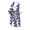
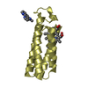
 Components Components | CYTOCHROME B562 | |||||||||
 Keywords Keywords | ELECTRON TRANSPORT | |||||||||
| Function / homology |  Function and homology information Function and homology informationelectron transport chain / periplasmic space / electron transfer activity / iron ion binding / heme binding Similarity search - Function | |||||||||
| Biological species |  | |||||||||
| Method |  X-RAY DIFFRACTION / Resolution: 1.4 Å X-RAY DIFFRACTION / Resolution: 1.4 Å | |||||||||
 Authors Authors | Hamada, K. / Bethge, P.H. / Mathews, F.S. | |||||||||
 Citation Citation | Journal: J.Mol.Biol. / Year: 1981 Title: Improvement of the 2.5 A resolution model of cytochrome b562 by redetermining the primary structure and using molecular graphics. Authors: Lederer, F. / Glatigny, A. / Bethge, P.H. / Bellamy, H.D. / Matthew, F.S. #1: Journal: J.Biol.Chem. / Year: 1979Title: The Structure of Cytochrome B562 from Escherichia Coli at 2.5 Angstroms Resolution Authors: Mathews, F.S. / Bethge, P.H. / Czerwinski, E.W. #2: Journal: J.Mol.Biol. / Year: 1974Title: Location of the Iron Atom and the Non-Crystallographic Symmetry Elements in Cytochrome B562 Authors: Czerwinski, E.W. / Mathews, F.S. | |||||||||
| History |
|
- Structure visualization
| Structure viewer | Molecule: MolmilJmol/JSmol |
|---|
- Downloads & links
-Download
| PDBx/mmCIF format | 256b.cif.gz | 58.6 KB | Display | PDBx/mmCIF format |
|---|---|---|---|---|
| PDB format | pdb256b.ent.gz | 43.2 KB | Display | PDB format |
| PDBx/mmJSON format | 256b.json.gz | Tree view | PDBx/mmJSON format | |
| Others |  Other downloads Other downloads |
-Validation report
| Arichive directory | https://data.pdbj.org/pub/pdb/validation_reports/56/256bftp://data.pdbj.org/pub/pdb/validation_reports/56/256b | HTTPS FTP |
|---|
-Related structure data
| Similar structure data |
|---|


-Links
 PDBj
PDBj


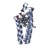
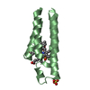
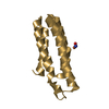
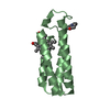
- Assembly
| Deposited unit | 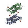
| ||||||||
|---|---|---|---|---|---|---|---|---|---|
| 1 | 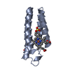
| ||||||||
| 2 | 
| ||||||||
| Unit cell |
| ||||||||
| Noncrystallographic symmetry (NCS) | NCS oper: (Code: given Matrix: (-0.0924, 0.21325, -0.97262), |
-Components
| #1: Protein | Mass: 11799.241 Da / Num. of mol.: 2 Source method: isolated from a genetically manipulated source Source: (gene. exp.) #2: Chemical | ChemComp-SO4 /   Mass: 96.063 Da / Num. of mol.: 4 / Source method: obtained synthetically / Formula: SO4 Mass: 96.063 Da / Num. of mol.: 4 / Source method: obtained synthetically / Formula: SO4#3: Chemical |   Mass: 616.487 Da / Num. of mol.: 2 / Source method: obtained synthetically / Formula: C34H32FeN4O4 Mass: 616.487 Da / Num. of mol.: 2 / Source method: obtained synthetically / Formula: C34H32FeN4O4#4: Water | ChemComp-HOH / |  Mass: 18.015 Da / Num. of mol.: 165 / Source method: isolated from a natural source / Formula: H2O Mass: 18.015 Da / Num. of mol.: 165 / Source method: isolated from a natural source / Formula: H2O |
|---|
-Experimental details
-Experiment
| Experiment | Method: X-RAY DIFFRACTION |
|---|
- Sample preparation
| Crystal | Density Matthews: 2.2 Å3/Da / Density % sol: 44.19 % | ||||||||||||||||||
|---|---|---|---|---|---|---|---|---|---|---|---|---|---|---|---|---|---|---|---|
| Crystal grow | *PLUS pH: 7.3 / Method: vapor diffusion / Details: Lederer, F., (1981) J. Mol. Biol., 148, 427. | ||||||||||||||||||
| Components of the solutions | *PLUS
|
-Data collection
| Radiation | Scattering type: x-ray |
|---|---|
| Radiation wavelength | Relative weight: 1 |
- Processing
| Software | Name: PROFFT / Classification: refinement | |||||||||||||||||||||||||||||||||||||||||||||||||||||||||||||||
|---|---|---|---|---|---|---|---|---|---|---|---|---|---|---|---|---|---|---|---|---|---|---|---|---|---|---|---|---|---|---|---|---|---|---|---|---|---|---|---|---|---|---|---|---|---|---|---|---|---|---|---|---|---|---|---|---|---|---|---|---|---|---|---|---|
| Refinement | Resolution: 1.4→6 Å / Rfactor obs: 0.164 | |||||||||||||||||||||||||||||||||||||||||||||||||||||||||||||||
| Refinement step | Cycle: LAST / Resolution: 1.4→6 Å
| |||||||||||||||||||||||||||||||||||||||||||||||||||||||||||||||
| Refine LS restraints |
|
