 Movie
Movie Controller


+ Open data

- Basic information
| Entry | Database: PDB / ID: 1g0b | ||||||
|---|---|---|---|---|---|---|---|
| Title | CARBONMONOXY LIGANDED EQUINE HEMOGLOBIN PH 8.5 | ||||||
 Components Components |
| ||||||
 Keywords Keywords | OXYGEN STORAGE/TRANSPORT / equine / hemoglobin / liganded / carbonmonoxy / protoporphyrin IX / OXYGEN STORAGE-TRANSPORT COMPLEX | ||||||
| Function / homology |  Function and homology information Function and homology informationhemoglobin alpha binding / haptoglobin-hemoglobin complex / hemoglobin complex / oxygen transport / erythrocyte development / oxygen carrier activity / oxygen binding / iron ion binding / heme binding / metal ion binding Similarity search - Function | ||||||
| Biological species |  | ||||||
| Method |  X-RAY DIFFRACTION / Resolution: 1.9 Å X-RAY DIFFRACTION / Resolution: 1.9 Å | ||||||
 Authors Authors | Mueser, T.C. / Rogers, P.H. / Arnone, A. | ||||||
 Citation Citation | Journal: Biochemistry / Year: 2000 Title: Interface sliding as illustrated by the multiple quaternary structures of liganded hemoglobin. Authors: Mueser, T.C. / Rogers, P.H. / Arnone, A. #1: Journal: J.Mol.Biol. / Year: 1977Title: The structure of horse methaemoglobin at 2-0 A resolution. Authors: Ladner, R.C. / Heidner, E.J. / Perutz, M.F. | ||||||
| History |
| ||||||
| Remark 999 | SEQUENCE The sequence is the same as in pdb entry 2MHB. This 1g0b structure is a re-refined ...SEQUENCE The sequence is the same as in pdb entry 2MHB. This 1g0b structure is a re-refined structure of R.C.LADNER,E.G.HEIDNER,M.F.PERUTZ from 1977. |
- Structure visualization
| Structure viewer | Molecule: MolmilJmol/JSmol |
|---|
- Downloads & links
-Download
| PDBx/mmCIF format | 1g0b.cif.gz | 70.8 KB | Display | PDBx/mmCIF format |
|---|---|---|---|---|
| PDB format | pdb1g0b.ent.gz | 53.5 KB | Display | PDB format |
| PDBx/mmJSON format | 1g0b.json.gz | Tree view | PDBx/mmJSON format | |
| Others |  Other downloads Other downloads |
-Validation report
| Arichive directory | https://data.pdbj.org/pub/pdb/validation_reports/g0/1g0bftp://data.pdbj.org/pub/pdb/validation_reports/g0/1g0b | HTTPS FTP |
|---|
-Related structure data


-Links
 PDBj
PDBj


- Assembly
| Deposited unit | 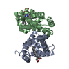
| ||||||||
|---|---|---|---|---|---|---|---|---|---|
| 1 | 
| ||||||||
| Unit cell |
| ||||||||
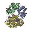
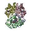
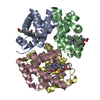
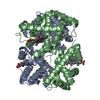
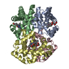
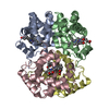
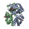
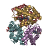
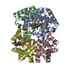
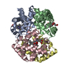
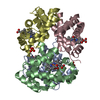
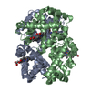
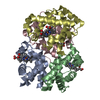
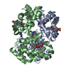
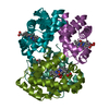
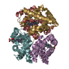
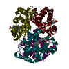
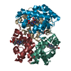
| Details | The biological assembly is a tetramer constructed from the heterodimer of chain A (alpha) and chain B (beta) and a symmetry related heterodimer of chain A (alpha) and chain B (beta) |
-Components
| #1: Protein | Mass: 15138.280 Da / Num. of mol.: 1 / Source method: isolated from a natural source / Source: (natural) | ||||||
|---|---|---|---|---|---|---|---|
| #2: Protein | Mass: 16032.274 Da / Num. of mol.: 1 / Source method: isolated from a natural source / Source: (natural) | ||||||
| #3: Chemical |   Mass: 616.487 Da / Num. of mol.: 2 / Source method: obtained synthetically / Formula: C34H32FeN4O4 Mass: 616.487 Da / Num. of mol.: 2 / Source method: obtained synthetically / Formula: C34H32FeN4O4#4: Chemical |   Mass: 28.010 Da / Num. of mol.: 2 / Source method: obtained synthetically / Formula: CO Mass: 28.010 Da / Num. of mol.: 2 / Source method: obtained synthetically / Formula: CO#5: Water | ChemComp-HOH / |  Mass: 18.015 Da / Num. of mol.: 72 / Source method: isolated from a natural source / Formula: H2O Mass: 18.015 Da / Num. of mol.: 72 / Source method: isolated from a natural source / Formula: H2OHas protein modification | N | |
-Experimental details
-Experiment
| Experiment | Method: X-RAY DIFFRACTION / Number of used crystals: 1 |
|---|
- Sample preparation
| Crystal | Density Matthews: 2.82 Å3/Da / Density % sol: 56.33 % / Description: AUTHOR USED THE SF DATA FROM ENTRY 2MHB. | |||||||||||||||
|---|---|---|---|---|---|---|---|---|---|---|---|---|---|---|---|---|
| Crystal grow | *PLUS | |||||||||||||||
| Components of the solutions | *PLUS
|
-Data collection
| Radiation | Scattering type: x-ray |
|---|---|
| Radiation wavelength | Relative weight: 1 |
| Reflection | *PLUS Num. measured all: 316145 |
| Reflection shell | *PLUS % possible obs: 87.8 % |
- Processing
| Software |
| ||||||||||||||||
|---|---|---|---|---|---|---|---|---|---|---|---|---|---|---|---|---|---|
| Refinement | Resolution: 1.9→8 Å / σ(F): 2 / σ(I): 0 / Stereochemistry target values: Engh & Huber
| ||||||||||||||||
| Refinement step | Cycle: LAST / Resolution: 1.9→8 Å
| ||||||||||||||||
| Refine LS restraints |
| ||||||||||||||||
| Software | *PLUS Name: PROLSQ / Classification: refinement | ||||||||||||||||
| Refinement | *PLUS | ||||||||||||||||
| Solvent computation | *PLUS | ||||||||||||||||
| Displacement parameters | *PLUS Biso mean: 24.8 Å2 |
