 Movie
Movie Controller

[English] 日本語
 Yorodumi
- PDB-1qpw: CRYSTAL STRUCTURE DETERMINATION OF PORCINE HEMOGLOBIN AT 1.8A RES... -
+ Open data

- Basic information
| Entry | Database: PDB / ID: 1qpw | ||||||
|---|---|---|---|---|---|---|---|
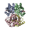
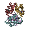
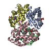
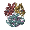
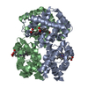
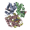
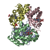
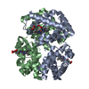
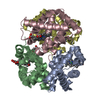
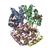
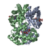
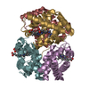
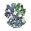
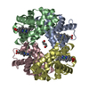
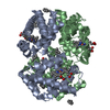
| Title | CRYSTAL STRUCTURE DETERMINATION OF PORCINE HEMOGLOBIN AT 1.8A RESOLUTION | ||||||
 Components Components |
| ||||||
 Keywords Keywords | OXYGEN TRANSPORT / X-RAY STUDY / PORCINE HEMOGLOBIN / ARTIFICIAL HUMAN BLOOD | ||||||
| Function / homology |  Function and homology information Function and homology informationhemoglobin alpha binding / haptoglobin-hemoglobin complex / hemoglobin complex / oxygen transport / erythrocyte development / oxygen carrier activity / oxygen binding / iron ion binding / heme binding / metal ion binding Similarity search - Function | ||||||
| Biological species |  | ||||||
| Method |  X-RAY DIFFRACTION / Resolution: 1.8 Å X-RAY DIFFRACTION / Resolution: 1.8 Å | ||||||
 Authors Authors | Lu, T.-H. / Panneerselvam, K. / Liaw, Y.-C. / Kan, P. / Lee, C.-J. | ||||||
 Citation Citation | Journal: Acta Crystallogr.,Sect.D / Year: 2000 Title: Structure determination of porcine haemoglobin. Authors: Lu, T.H. / Panneerselvam, K. / Liaw, Y.C. / Kan, P. / Lee, C.J. #1: Journal: J.Mol.Biol. / Year: 1994Title: Structure Determination of Aquomet Porcine Hemoglobin at 2.8A Resolution Authors: Katz, D.S. / White, S.P. / Huang, W. / Kumar, R. / Christianson, D.W. | ||||||
| History |
|
- Structure visualization
| Structure viewer | Molecule: MolmilJmol/JSmol |
|---|
- Downloads & links
-Download
| PDBx/mmCIF format | 1qpw.cif.gz | 139.8 KB | Display | PDBx/mmCIF format |
|---|---|---|---|---|
| PDB format | pdb1qpw.ent.gz | 108.8 KB | Display | PDB format |
| PDBx/mmJSON format | 1qpw.json.gz | Tree view | PDBx/mmJSON format | |
| Others |  Other downloads Other downloads |
-Validation report
| Arichive directory | https://data.pdbj.org/pub/pdb/validation_reports/qp/1qpwftp://data.pdbj.org/pub/pdb/validation_reports/qp/1qpw | HTTPS FTP |
|---|
-Related structure data
| Similar structure data |
|---|


-Links
 PDBj
PDBj


- Assembly
| Deposited unit | 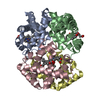
| ||||||||
|---|---|---|---|---|---|---|---|---|---|
| 1 |
| ||||||||
| Unit cell |
|
-Components
| #1: Protein | Mass: 15064.144 Da / Num. of mol.: 2 / Source method: isolated from a natural source / Details: TAIWANESE PIG FROM SLAUGHTERHOUSE / Source: (natural) #2: Protein | Mass: 16059.345 Da / Num. of mol.: 2 / Source method: isolated from a natural source / Details: TAIWANESE PIG FROM SLAUGHTERHOUSE / Source: (natural) #3: Chemical | ChemComp-HEM /   Mass: 616.487 Da / Num. of mol.: 4 / Source method: obtained synthetically / Formula: C34H32FeN4O4 Mass: 616.487 Da / Num. of mol.: 4 / Source method: obtained synthetically / Formula: C34H32FeN4O4#4: Chemical |   Mass: 31.999 Da / Num. of mol.: 2 / Source method: obtained synthetically / Formula: O2 Mass: 31.999 Da / Num. of mol.: 2 / Source method: obtained synthetically / Formula: O2#5: Water | ChemComp-HOH / |  Mass: 18.015 Da / Num. of mol.: 574 / Source method: isolated from a natural source / Formula: H2O Mass: 18.015 Da / Num. of mol.: 574 / Source method: isolated from a natural source / Formula: H2O |
|---|
-Experimental details
-Experiment
| Experiment | Method: X-RAY DIFFRACTION / Number of used crystals: 1 |
|---|
- Sample preparation
| Crystal | Density Matthews: 2.27 Å3/Da / Density % sol: 45.8 % | |||||||||||||||
|---|---|---|---|---|---|---|---|---|---|---|---|---|---|---|---|---|
| Crystal grow | Temperature: 296 K / Method: vapor diffusion, sitting drop / pH: 6.8 Details: 2.8M OF PHOSPHATE SOLUTION, pH 6.8, VAPOR DIFFUSION, SITTING DROP, temperature 296K | |||||||||||||||
| Crystal grow | *PLUS Method: vapor diffusion, hanging drop | |||||||||||||||
| Components of the solutions | *PLUS
|
-Data collection
| Diffraction | Mean temperature: 293 K |
|---|---|
| Diffraction source | Source: ROTATING ANODE / Type: RIGAKU RU300 / Wavelength: 1.5418 |
| Detector | Type: RIGAKU RAXIS / Detector: IMAGE PLATE / Date: Aug 10, 1991 |
| Radiation | Protocol: SINGLE WAVELENGTH / Monochromatic (M) / Laue (L): M / Scattering type: x-ray |
| Radiation wavelength | Wavelength: 1.5418 Å / Relative weight: 1 |
| Reflection | Resolution: 1.8→8 Å / Num. all: 37481 / Num. obs: 36820 / % possible obs: 78 % / Observed criterion σ(F): 2 / Observed criterion σ(I): 1 / Rmerge(I) obs: 0.058 / Net I/σ(I): 1 |
| Reflection shell | Resolution: 1.8→8 Å / Rmerge(I) obs: 0.058 / % possible all: 78 |
| Reflection | *PLUS Num. obs: 37481 |
| Reflection shell | *PLUS % possible obs: 78 % |
- Processing
| Software |
| ||||||||||||||||||||||||||||||||||||||||||||||||||||||||||||
|---|---|---|---|---|---|---|---|---|---|---|---|---|---|---|---|---|---|---|---|---|---|---|---|---|---|---|---|---|---|---|---|---|---|---|---|---|---|---|---|---|---|---|---|---|---|---|---|---|---|---|---|---|---|---|---|---|---|---|---|---|---|
| Refinement | Resolution: 1.8→8 Å / σ(F): 2 / σ(I): 1
| ||||||||||||||||||||||||||||||||||||||||||||||||||||||||||||
| Refinement step | Cycle: LAST / Resolution: 1.8→8 Å
| ||||||||||||||||||||||||||||||||||||||||||||||||||||||||||||
| Refine LS restraints |
| ||||||||||||||||||||||||||||||||||||||||||||||||||||||||||||
| Software | *PLUS Name: X-PLOR / Version: 3.1 / Classification: refinement | ||||||||||||||||||||||||||||||||||||||||||||||||||||||||||||
| Refinement | *PLUS | ||||||||||||||||||||||||||||||||||||||||||||||||||||||||||||
| Solvent computation | *PLUS | ||||||||||||||||||||||||||||||||||||||||||||||||||||||||||||
| Displacement parameters | *PLUS Biso mean: 34.8 Å2 | ||||||||||||||||||||||||||||||||||||||||||||||||||||||||||||
| Refine LS restraints | *PLUS
|
