 Movie
Movie Controller


+ Open data

- Basic information
| Entry | Database: PDB / ID: 1g0a | ||||||
|---|---|---|---|---|---|---|---|
| Title | CARBONMONOXY LIGANDED BOVINE HEMOGLOBIN PH 8.5 | ||||||
 Components Components |
| ||||||
 Keywords Keywords | OXYGEN STORAGE/TRANSPORT / bovine / hemoglobin / liganded / carbonmonoxy / protoporphyrin IX / OXYGEN STORAGE-TRANSPORT COMPLEX | ||||||
| Function / homology |  Function and homology information Function and homology informationScavenging of heme from plasma / Heme signaling / Erythrocytes take up carbon dioxide and release oxygen / Erythrocytes take up oxygen and release carbon dioxide / Cytoprotection by HMOX1 / Neutrophil degranulation / hemoglobin alpha binding / haptoglobin-hemoglobin complex / hemoglobin complex / oxygen transport ...Scavenging of heme from plasma / Heme signaling / Erythrocytes take up carbon dioxide and release oxygen / Erythrocytes take up oxygen and release carbon dioxide / Cytoprotection by HMOX1 / Neutrophil degranulation / hemoglobin alpha binding / haptoglobin-hemoglobin complex / hemoglobin complex / oxygen transport / erythrocyte development / oxygen carrier activity / oxygen binding / iron ion binding / heme binding / metal ion binding Similarity search - Function | ||||||
| Biological species |  | ||||||
| Method |  X-RAY DIFFRACTION / Resolution: 2.04 Å X-RAY DIFFRACTION / Resolution: 2.04 Å | ||||||
 Authors Authors | Mueser, T.C. / Rogers, P.H. / Arnone, A. | ||||||
 Citation Citation | Journal: Biochemistry / Year: 2000 Title: Interface sliding as illustrated by the multiple quaternary structures of liganded hemoglobin. Authors: Mueser, T.C. / Rogers, P.H. / Arnone, A. | ||||||
| History |
|
- Structure visualization
| Structure viewer | Molecule: MolmilJmol/JSmol |
|---|
- Downloads & links
-Download
| PDBx/mmCIF format | 1g0a.cif.gz | 127.4 KB | Display | PDBx/mmCIF format |
|---|---|---|---|---|
| PDB format | pdb1g0a.ent.gz | 101 KB | Display | PDB format |
| PDBx/mmJSON format | 1g0a.json.gz | Tree view | PDBx/mmJSON format | |
| Others |  Other downloads Other downloads |
-Validation report
| Arichive directory | https://data.pdbj.org/pub/pdb/validation_reports/g0/1g0aftp://data.pdbj.org/pub/pdb/validation_reports/g0/1g0a | HTTPS FTP |
|---|
-Related structure data
-Links
 PDBj
PDBj


- Assembly
| Deposited unit | 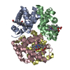
| ||||||||
|---|---|---|---|---|---|---|---|---|---|
| 1 |
| ||||||||
| Unit cell |
| ||||||||
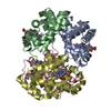
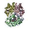
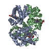
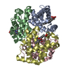
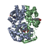
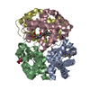
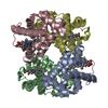
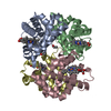
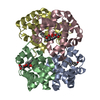
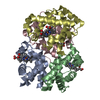
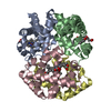
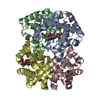
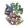
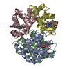
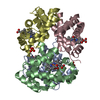
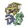
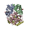
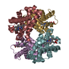
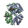
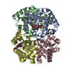
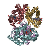
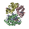
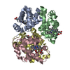
| Details | The biological assembly is a tetramer constructed from the heterodimer of chain A (alpha) and chain B (beta) and the heterodimer of chain C (alpha) and chain D (beta) |
-Components
| #1: Protein | Mass: 15077.171 Da / Num. of mol.: 2 / Source method: isolated from a natural source / Source: (natural) #2: Protein | Mass: 15977.382 Da / Num. of mol.: 2 / Source method: isolated from a natural source / Source: (natural) #3: Chemical | ChemComp-HEM /   Mass: 616.487 Da / Num. of mol.: 4 / Source method: obtained synthetically / Formula: C34H32FeN4O4 Mass: 616.487 Da / Num. of mol.: 4 / Source method: obtained synthetically / Formula: C34H32FeN4O4#4: Chemical | ChemComp-CMO /   Mass: 28.010 Da / Num. of mol.: 4 / Source method: obtained synthetically / Formula: CO Mass: 28.010 Da / Num. of mol.: 4 / Source method: obtained synthetically / Formula: CO#5: Water | ChemComp-HOH / |  Mass: 18.015 Da / Num. of mol.: 145 / Source method: isolated from a natural source / Formula: H2O Mass: 18.015 Da / Num. of mol.: 145 / Source method: isolated from a natural source / Formula: H2O |
|---|
-Experimental details
-Experiment
| Experiment | Method: X-RAY DIFFRACTION / Number of used crystals: 1 |
|---|
- Sample preparation
| Crystal | Density Matthews: 2.46 Å3/Da / Density % sol: 50.04 % | ||||||||||||||||||||||||||||||
|---|---|---|---|---|---|---|---|---|---|---|---|---|---|---|---|---|---|---|---|---|---|---|---|---|---|---|---|---|---|---|---|
| Crystal grow | Temperature: 298 K / Method: vapor diffusion, hanging drop / pH: 8.5 Details: sodium TAPS, ammonium sulphate, potassium chloride, polyethylene glycol 3350, pH 8.5, VAPOR DIFFUSION, HANGING DROP, temperature 298K | ||||||||||||||||||||||||||||||
| Crystal grow | *PLUS | ||||||||||||||||||||||||||||||
| Components of the solutions | *PLUS
|
-Data collection
| Diffraction | Mean temperature: 298 K |
|---|---|
| Diffraction source | Source: ROTATING ANODE / Type: RIGAKU RU200 / Wavelength: 1.5418 |
| Detector | Type: SDMS / Detector: AREA DETECTOR / Date: Jan 15, 1992 |
| Radiation | Protocol: SINGLE WAVELENGTH / Monochromatic (M) / Laue (L): M / Scattering type: x-ray |
| Radiation wavelength | Wavelength: 1.5418 Å / Relative weight: 1 |
| Reflection | Resolution: 2.04→35.53 Å / Num. all: 39833 / Num. obs: 38330 / % possible obs: 96.2 % / Observed criterion σ(F): 0 / Observed criterion σ(I): 0 / Redundancy: 7.95 % / Biso Wilson estimate: 29.2 Å2 / Rmerge(I) obs: 0.067 / Net I/σ(I): 12.9 |
| Reflection shell | Resolution: 2.04→2.2 Å / Redundancy: 3.1 % / Rmerge(I) obs: 0.197 / Num. unique all: 6759 / % possible all: 86.4 |
| Reflection | *PLUS Num. measured all: 304865 |
| Reflection shell | *PLUS % possible obs: 86.4 % |
- Processing
| Software |
| ||||||||||||||||
|---|---|---|---|---|---|---|---|---|---|---|---|---|---|---|---|---|---|
| Refinement | Resolution: 2.04→8 Å / σ(F): 2 / σ(I): 0 / Stereochemistry target values: Engh & Huber
| ||||||||||||||||
| Refinement step | Cycle: LAST / Resolution: 2.04→8 Å
| ||||||||||||||||
| Refine LS restraints |
| ||||||||||||||||
| Software | *PLUS Name: PROLSQ / Classification: refinement | ||||||||||||||||
| Refinement | *PLUS | ||||||||||||||||
| Solvent computation | *PLUS | ||||||||||||||||
| Displacement parameters | *PLUS Biso mean: 26.9 Å2 |
