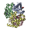
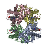
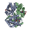
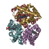
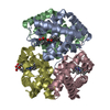
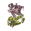
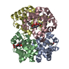
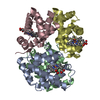
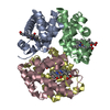
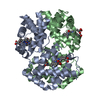
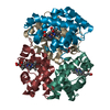
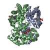
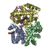
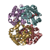
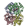
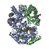
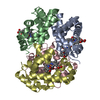
- PDB-6ii1: Crystal Structure Analysis of CO form hemoglobin from Bos taurus -
+
Open data
ID or keywords:
Loading...
-
Basic information
Entry
Database: PDB / ID: 6ii1
Title
Crystal Structure Analysis of CO form hemoglobin from Bos taurus
Components
Hemoglobin subunit alpha
Hemoglobin subunit beta
Keywords
OXYGEN TRANSPORT / heme binding / iron ion binding / Red blood cells / globin family
Function / homology
Function and homology information
Scavenging of heme from plasma / Heme signaling / Erythrocytes take up carbon dioxide and release oxygen / Erythrocytes take up oxygen and release carbon dioxide / Cytoprotection by HMOX1 / Neutrophil degranulation / hemoglobin alpha binding / haptoglobin-hemoglobin complex / hemoglobin complex / oxygen transport ...Scavenging of heme from plasma / Heme signaling / Erythrocytes take up carbon dioxide and release oxygen / Erythrocytes take up oxygen and release carbon dioxide / Cytoprotection by HMOX1 / Neutrophil degranulation / hemoglobin alpha binding / haptoglobin-hemoglobin complex / hemoglobin complex / oxygen transport / erythrocyte development / oxygen carrier activity / oxygen binding / iron ion binding / heme binding / metal ion binding Similarity search - Function
In the structure databanks used in Yorodumi, some data are registered as the other names, "COVID-19 virus" and "2019-nCoV". Here are the details of the virus and the list of structure data.
Jan 31, 2019. EMDB accession codes are about to change! (news from PDBe EMDB page)
EMDB accession codes are about to change! (news from PDBe EMDB page)
The allocation of 4 digits for EMDB accession codes will soon come to an end. Whilst these codes will remain in use, new EMDB accession codes will include an additional digit and will expand incrementally as the available range of codes is exhausted. The current 4-digit format prefixed with “EMD-” (i.e. EMD-XXXX) will advance to a 5-digit format (i.e. EMD-XXXXX), and so on. It is currently estimated that the 4-digit codes will be depleted around Spring 2019, at which point the 5-digit format will come into force.
The EM Navigator/Yorodumi systems omit the EMD- prefix.
Related info.:Q: What is EMD? / ID/Accession-code notation in Yorodumi/EM Navigator
Yorodumi is a browser for structure data from EMDB, PDB, SASBDB, etc.
This page is also the successor to EM Navigator detail page, and also detail information page/front-end page for Omokage search.
The word "yorodu" (or yorozu) is an old Japanese word meaning "ten thousand". "mi" (miru) is to see.
Related info.:EMDB / PDB / SASBDB / Comparison of 3 databanks / Yorodumi Search / Aug 31, 2016. New EM Navigator & Yorodumi / Yorodumi Papers / Jmol/JSmol / Function and homology information / Changes in new EM Navigator and Yorodumi
 Movie
Movie Controller


 Open data
 Basic information
 Components
 Keywords
 Function and homology information
 X-RAY DIFFRACTION /
  Authors
 Citation
 Structure visualization
 Downloads & links
 Other downloads
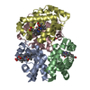

 PDBj
PDBj


 Assembly
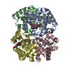

 Mass: 616.487 Da / Num. of mol.: 4 / Source method: obtained synthetically / Formula: C34H32FeN4O4
 Mass: 28.010 Da / Num. of mol.: 4 / Source method: obtained synthetically / Formula: CO
 Mass: 18.015 Da / Num. of mol.: 723 / Source method: isolated from a natural source / Formula: H2O
 Sample preparation
 / Beamline: AR-NW12A / Wavelength: 1 Å
 Processing