 Movie
Movie Controller

[English] 日本語
 Yorodumi
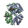
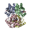
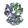
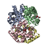
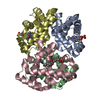
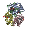
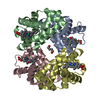
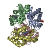
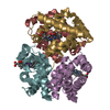
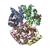
- PDB-2mhb: THE STRUCTURE OF HORSE METHAEMOGLOBIN AT 2.0 ANGSTROMS RESOLUTION -
+ Open data

- Basic information
| Entry | Database: PDB / ID: 2mhb | |||||||||
|---|---|---|---|---|---|---|---|---|---|---|
| Title | THE STRUCTURE OF HORSE METHAEMOGLOBIN AT 2.0 ANGSTROMS RESOLUTION | |||||||||
 Components Components |
| |||||||||
 Keywords Keywords | OXYGEN TRANSPORT | |||||||||
| Function / homology |  Function and homology information Function and homology informationhemoglobin alpha binding / haptoglobin-hemoglobin complex / hemoglobin complex / oxygen transport / erythrocyte development / oxygen carrier activity / oxygen binding / iron ion binding / heme binding / metal ion binding Similarity search - Function | |||||||||
| Biological species |  | |||||||||
| Method |  X-RAY DIFFRACTION / Resolution: 2 Å X-RAY DIFFRACTION / Resolution: 2 Å | |||||||||
 Authors Authors | Ladner, R.C. / Heidner, E.G. / Perutz, M.F. | |||||||||
 Citation Citation | Journal: J.Mol.Biol. / Year: 1977 Title: The structure of horse methaemoglobin at 2-0 A resolution. Authors: Ladner, R.C. / Heidner, E.J. / Perutz, M.F. #1: Journal: Nature / Year: 1968Title: Three-Dimensional Fourier Synthesis of Horse Oxyhaemoglobin at 2.8 Angstroms Resolution,the Atomic Model Authors: Perutz, M.F. / Muirhead, H. / Cox, J.M. / Goaman, L.C.G. #2: Journal: J.Mol.Biol. / Year: 1976Title: A Correction to the Sequence of the Alpha Chains of Horse Haemoglobin Authors: Ladner, R.C. / Air, G.M. / Fogg, J.H. | |||||||||
| History |
|
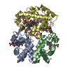
- Structure visualization
| Structure viewer | Molecule: MolmilJmol/JSmol |
|---|
- Downloads & links
-Download
| PDBx/mmCIF format | 2mhb.cif.gz | 69.6 KB | Display | PDBx/mmCIF format |
|---|---|---|---|---|
| PDB format | pdb2mhb.ent.gz | 52.6 KB | Display | PDB format |
| PDBx/mmJSON format | 2mhb.json.gz | Tree view | PDBx/mmJSON format | |
| Others |  Other downloads Other downloads |
-Validation report
| Arichive directory | https://data.pdbj.org/pub/pdb/validation_reports/mh/2mhbftp://data.pdbj.org/pub/pdb/validation_reports/mh/2mhb | HTTPS FTP |
|---|
-Related structure data
| Similar structure data |
|---|


-Links
 PDBj
PDBj


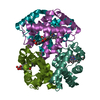
- Assembly
| Deposited unit | 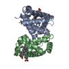
| ||||||||
|---|---|---|---|---|---|---|---|---|---|
| 1 | 
| ||||||||
| Unit cell |
|
-Components
| #1: Protein | Mass: 15138.280 Da / Num. of mol.: 1 Source method: isolated from a genetically manipulated source Source: (gene. exp.) | ||||
|---|---|---|---|---|---|
| #2: Protein | Mass: 16032.274 Da / Num. of mol.: 1 Source method: isolated from a genetically manipulated source Source: (gene. exp.) | ||||
| #3: Chemical |   Mass: 616.487 Da / Num. of mol.: 2 / Source method: obtained synthetically / Formula: C34H32FeN4O4 Mass: 616.487 Da / Num. of mol.: 2 / Source method: obtained synthetically / Formula: C34H32FeN4O4#4: Water | ChemComp-HOH / |  Mass: 18.015 Da / Num. of mol.: 2 / Source method: isolated from a natural source / Formula: H2O Mass: 18.015 Da / Num. of mol.: 2 / Source method: isolated from a natural source / Formula: H2OHas protein modification | N | |
-Experimental details
-Experiment
| Experiment | Method: X-RAY DIFFRACTION |
|---|
- Sample preparation
| Crystal | Density Matthews: 2.79 Å3/Da / Density % sol: 55.95 % |
|---|---|
| Crystal grow | *PLUS Method: other / Details: Perutz, M.F., (1968) J. Crystal Growth, 2, 54. |
-Data collection
| Radiation | Scattering type: x-ray |
|---|---|
| Radiation wavelength | Relative weight: 1 |
| Reflection | *PLUS Num. obs: 23962 |
- Processing
| Refinement | Rfactor Rwork: 0.23 / Rfactor obs: 0.23 / Highest resolution: 2 Å Details: RESIDUES LYS A 61 AND LYS B 66 MAY FORM HYDROGEN BONDS TO THE PROPIONIC ACID SIDE CHAINS OF THEIR RESPECTIVE HEME GROUPS BUT THESE REGIONS ARE POORLY RESOLVED. | ||||||||||||
|---|---|---|---|---|---|---|---|---|---|---|---|---|---|
| Refinement step | Cycle: LAST / Highest resolution: 2 Å
| ||||||||||||
| Refinement | *PLUS Rfactor obs: 0.231 | ||||||||||||
| Solvent computation | *PLUS | ||||||||||||
| Displacement parameters | *PLUS |
