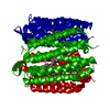
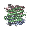
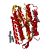
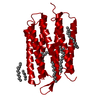
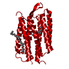
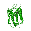
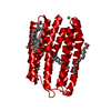
ジャーナル: Nature / 年: 2000 タイトル: Molecular mechanism of vectorial proton translocation by bacteriorhodopsin. 著者: S Subramaniam / R Henderson / 要旨: Bacteriorhodopsin, a membrane protein with a relative molecular mass of 27,000, is a light driven pump which transports protons across the cell membrane of the halophilic organism Halobacterium ...Bacteriorhodopsin, a membrane protein with a relative molecular mass of 27,000, is a light driven pump which transports protons across the cell membrane of the halophilic organism Halobacterium salinarum. The chromophore retinal is covalently attached to the protein via a protonated Schiff base. Upon illumination, retinal is isomerized. The Schiff base then releases a proton to the extracellular medium, and is subsequently reprotonated from the cytoplasm. An atomic model for bacteriorhodopsin was first determined by Henderson et al, and has been confirmed and extended by work in a number of laboratories in the last few years. Here we present an atomic model for structural changes involved in the vectorial, light-driven transport of protons by bacteriorhodopsin. A 'switch' mechanism ensures the vectorial nature of pumping. First, retinal unbends, triggered by loss of the Schiff base proton, and second, a protein conformational change occurs. This conformational change, which we have determined by electron crystallography at atomic (3.2 A in-plane and 3.6 A vertical) resolution, is largely localized to helices F and G, and provides an 'opening' of the protein to protons on the cytoplasmic side of the membrane.
履歴
登録
2000年7月15日
登録サイト: RCSB / 処理サイト: RCSB
改定 1.0
2000年8月9日
Provider: repository / タイプ: Initial release
改定 1.1
2008年4月27日
Group: Version format compliance
改定 1.2
2011年7月13日
Group: Derived calculations / Version format compliance
温度: 310 K / 手法: naturally occurring in vivo / pH: 7 詳細: crystal size is increased by fusion and annealing using detergents, pH 7, naturally occurring in vivo, temperature 37K
結晶化
*PLUS
温度: 4 ℃ / pH: 5.6 / 手法: unknown
溶液の組成
*PLUS
ID
濃度
一般名
Crystal-ID
Sol-ID
1
18-23 mg/ml
protein
1
1
2
0.5 %(w/v)
beta-octylglucopyranoside
1
1
3
4 %(w/v)
benzamidine
1
1
4
1.75M
sodiumphosphate
1
1
5
1.8-2.3 M
ammoniumsulfate
1
reservoir
-
データ収集
EM imaging
Specimen-ID: 1
ID
加速電圧 (kV)
詳細
照射モード
モデル
モード
最高温度 (K)
凍結剤
倍率(公称値) (X)
電子線源
1
120
60degreetiltedspecimens
FLOODBEAM
FEI/PHILIPS EM420
DIFFRACTION
153
2
100
0, 20, 45degree + randomdegreetilts
FLOODBEAM
SIEMENS SULEIKA
BRIGHTFIELD
5
HELIUM
66000
3
100
, 20, 45degree + randomdegreetilts
SPOTSCAN
JEOL 100B
BRIGHTFIELD
158
NITROGEN
55000
FIELD EMISSION GUN
撮影
ID
Imaging-ID
平均露光時間 (sec.)
電子線照射量 (e/Å2)
フィルム・検出器のモデル
実像数
Num. of diffraction images
2
2
12
20
GENERIC FILM
52
3
3
15
GENERIC FILM
20
1
1
GENERIC FILM
150
回折
平均測定温度: 93 K
放射光源
由来: ELECTRON MICROSCOPE / タイプ: OTHER / 波長: 0.033
検出器
タイプ: OTHER / 検出器: FILM / 日付: 1986年1月1日
放射
プロトコル: SINGLE WAVELENGTH / 単色(M)・ラウエ(L): M / 散乱光タイプ: electron
解像度: 3.2→200 Å / 立体化学のターゲット値: Engh & Huber 詳細: For the tilt angles used, the maximal possible theoretical completeness of the data set is ~87%. The completeness of our data is close to this limit up to 3.5 Angstroms. The completeness ...詳細: For the tilt angles used, the maximal possible theoretical completeness of the data set is ~87%. The completeness of our data is close to this limit up to 3.5 Angstroms. The completeness drops to 65.1% when all of the data to 3.2 Angstroms is included.
 ムービー
ムービー コントローラー


 データを開く
 基本情報
基本情報 要素
要素 キーワード
キーワード 機能・相同性情報
 Halobacterium salinarum (好塩性)
 データ登録者
 引用
引用
 構造の表示
構造の表示 ダウンロードとリンク
 その他のダウンロード
 PDBj
PDBj


 集合体
集合体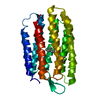

 分子量: 284.436 Da / 分子数: 1 / 由来タイプ: 合成 / 式: C20H28O
 試料調製
試料調製 FIELD EMISSION GUN
 解析
解析