 Movie
Movie Controller

[English] 日本語
 Yorodumi
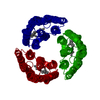
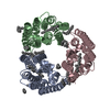
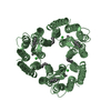
- PDB-6k6k: The crystal structure of light-driven cyanobacterial chloride imp... -
+ Open data

- Basic information
| Entry | Database: PDB / ID: 6k6k | |||||||||
|---|---|---|---|---|---|---|---|---|---|---|
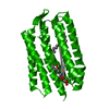
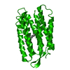
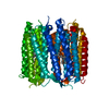
| Title | The crystal structure of light-driven cyanobacterial chloride importer (N63A/P118A) Mastigocladopsis repens | |||||||||
 Components Components | Cyanobacterial chloride importer | |||||||||
 Keywords Keywords | MEMBRANE PROTEIN / chloride ion pump / rhodopsin | |||||||||
| Function / homology | Rhopdopsin 7-helix transmembrane proteins / Rhodopsin 7-helix transmembrane proteins / Up-down Bundle / Mainly Alpha / OLEIC ACID / RETINAL Function and homology information Function and homology information | |||||||||
| Biological species |  Mastigocladopsis repens (bacteria) Mastigocladopsis repens (bacteria) | |||||||||
| Method |  X-RAY DIFFRACTION / SYNCHROTRON / MOLECULAR REPLACEMENT / Resolution: 2.197 Å X-RAY DIFFRACTION / SYNCHROTRON / MOLECULAR REPLACEMENT / Resolution: 2.197 Å | |||||||||
 Authors Authors | Yun, J.H. / Park, J.H. / Jin, Z. / Ohki, M. / Wang, Y. / Lupala, C.S. / Kim, M. / Liu, H. / Park, S.Y. / Lee, W. | |||||||||
| Funding support |  Korea, Republic Of, 2items Korea, Republic Of, 2items
| |||||||||
 Citation Citation | Journal: To Be Published Title: The crystal structure of light-driven cyanobacterial chloride importer (N63A/P118A) Mastigocladopsis repens Authors: Yun, J.H. / Park, J.H. / Jin, Z. / Ohki, M. / Wang, Y. / Lupala, C.S. / Kim, M. / Liu, H. / Park, S.Y. / Lee, W. | |||||||||
| History |
|
- Structure visualization
| Structure viewer | Molecule: MolmilJmol/JSmol |
|---|
- Downloads & links
-Download
| PDBx/mmCIF format | 6k6k.cif.gz | 61.4 KB | Display | PDBx/mmCIF format |
|---|---|---|---|---|
| PDB format | pdb6k6k.ent.gz | 43.2 KB | Display | PDB format |
| PDBx/mmJSON format | 6k6k.json.gz | Tree view | PDBx/mmJSON format | |
| Others |  Other downloads Other downloads |
-Validation report
| Arichive directory | https://data.pdbj.org/pub/pdb/validation_reports/k6/6k6kftp://data.pdbj.org/pub/pdb/validation_reports/k6/6k6k | HTTPS FTP |
|---|
-Related structure data
| Related structure data |  5itcS S: Starting model for refinement |
|---|---|
| Similar structure data |

-Links
 PDBj
PDBj


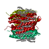
- Assembly
| Deposited unit | 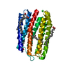
| ||||||||
|---|---|---|---|---|---|---|---|---|---|
| 1 | 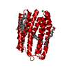
| ||||||||
| Unit cell |
| ||||||||
| Components on special symmetry positions |
|
-Components
| #1: Protein | Mass: 25991.463 Da / Num. of mol.: 1 / Mutation: N63A/P118A Source method: isolated from a genetically manipulated source Source: (gene. exp.) Mastigocladopsis repens (bacteria) / Production host: | ||||||||||||
|---|---|---|---|---|---|---|---|---|---|---|---|---|---|
| #2: Chemical |   Mass: 35.453 Da / Num. of mol.: 2 / Source method: obtained synthetically / Formula: Cl Mass: 35.453 Da / Num. of mol.: 2 / Source method: obtained synthetically / Formula: Cl#3: Chemical | ChemComp-RET / |   Mass: 284.436 Da / Num. of mol.: 1 / Source method: obtained synthetically / Formula: C20H28O / Feature type: SUBJECT OF INVESTIGATION Mass: 284.436 Da / Num. of mol.: 1 / Source method: obtained synthetically / Formula: C20H28O / Feature type: SUBJECT OF INVESTIGATION#4: Chemical | ChemComp-OLA /   Mass: 282.461 Da / Num. of mol.: 4 / Source method: obtained synthetically / Formula: C18H34O2 Mass: 282.461 Da / Num. of mol.: 4 / Source method: obtained synthetically / Formula: C18H34O2#5: Water | ChemComp-HOH / |  Mass: 18.015 Da / Num. of mol.: 52 / Source method: isolated from a natural source / Formula: H2O Mass: 18.015 Da / Num. of mol.: 52 / Source method: isolated from a natural source / Formula: H2OHas ligand of interest | Y | Has protein modification | Y | Sequence details | Residues N63A/P118A represent mutations. | |
-Experimental details
-Experiment
| Experiment | Method: X-RAY DIFFRACTION / Number of used crystals: 1 |
|---|
- Sample preparation
| Crystal | Density Matthews: 2.26 Å3/Da / Density % sol: 45.48 % |
|---|---|
| Crystal grow | Temperature: 293 K / Method: lipidic cubic phase / pH: 4.9 / Details: Magnesium nitrate hexahydrate, Sodium citrate |
-Data collection
| Diffraction | Mean temperature: 93 K / Serial crystal experiment: N |
|---|---|
| Diffraction source | Source: SYNCHROTRON / Site: PAL/PLS / Beamline: 5C (4A) / Wavelength: 0.987 Å |
| Detector | Type: ADSC QUANTUM 315r / Detector: CCD / Date: Nov 13, 2018 |
| Radiation | Protocol: SINGLE WAVELENGTH / Monochromatic (M) / Laue (L): M / Scattering type: x-ray |
| Radiation wavelength | Wavelength: 0.987 Å / Relative weight: 1 |
| Reflection | Resolution: 2.197→36.83 Å / Num. obs: 11542 / % possible obs: 93 % / Redundancy: 12.4 % / Net I/σ(I): 17.1 |
| Reflection shell | Resolution: 2.2→2.24 Å / Num. unique obs: 489 |
- Processing
| Software |
| |||||||||||||||||||||||||||||||||||
|---|---|---|---|---|---|---|---|---|---|---|---|---|---|---|---|---|---|---|---|---|---|---|---|---|---|---|---|---|---|---|---|---|---|---|---|---|
| Refinement | Method to determine structure: MOLECULAR REPLACEMENT Starting model: 5ITC Resolution: 2.197→36.829 Å / SU ML: 0.3 / Cross valid method: FREE R-VALUE / σ(F): 1.87 / Phase error: 23.65
| |||||||||||||||||||||||||||||||||||
| Solvent computation | Shrinkage radii: 0.9 Å / VDW probe radii: 1.11 Å | |||||||||||||||||||||||||||||||||||
| Refinement step | Cycle: LAST / Resolution: 2.197→36.829 Å
| |||||||||||||||||||||||||||||||||||
| Refine LS restraints |
| |||||||||||||||||||||||||||||||||||
| LS refinement shell |
|
