 Movie
Movie Controller

[English] 日本語
 Yorodumi
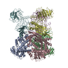
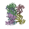
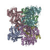
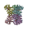
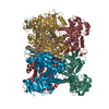
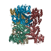
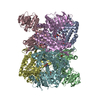
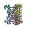
- PDB-1euz: GLUTAMATE DEHYDROGENASE FROM THERMOCOCCUS PROFUNDUS IN THE UNLIGA... -
+ Open data

- Basic information
| Entry | Database: PDB / ID: 1euz | ||||||
|---|---|---|---|---|---|---|---|
| Title | GLUTAMATE DEHYDROGENASE FROM THERMOCOCCUS PROFUNDUS IN THE UNLIGATED STATE | ||||||
 Components Components | GLUTAMATE DEHYDROGENASE | ||||||
 Keywords Keywords | OXIDOREDUCTASE / GLUTAMATE / HYPERTHERMOSTABILITY / domain closure movement | ||||||
| Function / homology |  Function and homology information Function and homology informationglutamate dehydrogenase [NAD(P)+] / L-glutamate dehydrogenase (NADP+) activity / L-glutamate dehydrogenase (NAD+) activity / L-glutamate catabolic process Similarity search - Function | ||||||
| Biological species |   Thermococcus profundus (archaea) Thermococcus profundus (archaea) | ||||||
| Method |  X-RAY DIFFRACTION / SYNCHROTRON / MOLECULAR REPLACEMENT / Resolution: 2.25 Å X-RAY DIFFRACTION / SYNCHROTRON / MOLECULAR REPLACEMENT / Resolution: 2.25 Å | ||||||
 Authors Authors | Nakasako, M. | ||||||
 Citation Citation | Journal: Biochemistry / Year: 2001 Title: Large-scale domain movements and hydration structure changes in the active-site cleft of unligated glutamate dehydrogenase from Thermococcus profundus studied by cryogenic X-ray crystal ...Title: Large-scale domain movements and hydration structure changes in the active-site cleft of unligated glutamate dehydrogenase from Thermococcus profundus studied by cryogenic X-ray crystal structure analysis and small-angle X-ray scattering. Authors: Nakasako, M. / Fujisawa, T. / Adachi, S. / Kudo, T. / Higuchi, S. #1: Journal: Acta Crystallogr.,Sect.D / Year: 1999Title: Crystallization and Preliminary X-ray Diffraction Studies of Hyperthermostable Glutamate Dehydrogenase from Thermococcus profundus Authors: Higuchi, S. / Nakasako, M. / Kudo, T. | ||||||
| History |
|
- Structure visualization
| Structure viewer | Molecule: MolmilJmol/JSmol |
|---|
- Downloads & links
-Download
| PDBx/mmCIF format | 1euz.cif.gz | 468.6 KB | Display | PDBx/mmCIF format |
|---|---|---|---|---|
| PDB format | pdb1euz.ent.gz | 390.2 KB | Display | PDB format |
| PDBx/mmJSON format | 1euz.json.gz | Tree view | PDBx/mmJSON format | |
| Others |  Other downloads Other downloads |
-Validation report
| Arichive directory | https://data.pdbj.org/pub/pdb/validation_reports/eu/1euzftp://data.pdbj.org/pub/pdb/validation_reports/eu/1euz | HTTPS FTP |
|---|
-Related structure data
| Related structure data | |
|---|---|
| Similar structure data |


-Links
 PDBj
PDBj- Assembly
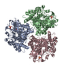
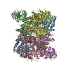
| Deposited unit | 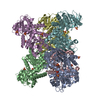
| ||||||||
|---|---|---|---|---|---|---|---|---|---|
| 1 |
| ||||||||
| Unit cell |
|
-Components
| #1: Protein | Mass: 46758.477 Da / Num. of mol.: 6 Source method: isolated from a genetically manipulated source Source: (gene. exp.) Thermococcus profundus (archaea) / Production host:  #2: Chemical | ChemComp-SO4 /   Mass: 96.063 Da / Num. of mol.: 17 / Source method: obtained synthetically / Formula: SO4 Mass: 96.063 Da / Num. of mol.: 17 / Source method: obtained synthetically / Formula: SO4 |
|---|
-Experimental details
-Experiment
| Experiment | Method: X-RAY DIFFRACTION / Number of used crystals: 1 |
|---|
- Sample preparation
| Crystal | Density Matthews: 3.97 Å3/Da / Density % sol: 69.03 % Description: DATA WERE COLLECTED USING THE OSCILLATION METHOD | |||||||||||||||||||||||||
|---|---|---|---|---|---|---|---|---|---|---|---|---|---|---|---|---|---|---|---|---|---|---|---|---|---|---|
| Crystal grow | Temperature: 293 K / Method: vapor diffusion, hanging drop / pH: 5 Details: PEG8000, Lithium sulfate, sodium acetate, pH 5.0, VAPOR DIFFUSION, HANGING DROP, temperature 293K | |||||||||||||||||||||||||
| Crystal grow | *PLUS | |||||||||||||||||||||||||
| Components of the solutions | *PLUS
|
-Data collection
| Diffraction | Mean temperature: 110 K |
|---|---|
| Diffraction source | Source: SYNCHROTRON / Site: SPring-8  / Beamline: BL44B2 / Wavelength: 1 / Beamline: BL44B2 / Wavelength: 1 |
| Detector | Type: RIGAKU RAXIS IV / Detector: IMAGE PLATE / Date: Mar 15, 1999 / Details: PT-COATED CYLINDRICAL BENT MIRROR |
| Radiation | Monochromator: FIXED EXIT DOUBLE CRYSTAL / Protocol: SINGLE WAVELENGTH / Monochromatic (M) / Laue (L): M / Scattering type: x-ray |
| Radiation wavelength | Wavelength: 1 Å / Relative weight: 1 |
| Reflection | Resolution: 2.25→100 Å / Num. all: 206382 / Num. obs: 206382 / % possible obs: 99.7 % / Observed criterion σ(F): 1488939 / Observed criterion σ(I): 1 / Redundancy: 7.21 % / Rmerge(I) obs: 0.072 / Net I/σ(I): 17.9 |
| Reflection shell | Resolution: 2.25→2.33 Å / Rmerge(I) obs: 0.31 / Mean I/σ(I) obs: 3.4 / % possible all: 99.4 |
| Reflection | *PLUS Num. measured all: 1488939 / Rmerge(I) obs: 0.063 |
| Reflection shell | *PLUS % possible obs: 99.4 % / Rmerge(I) obs: 0.322 |
- Processing
| Software |
| |||||||||||||||||||||||||
|---|---|---|---|---|---|---|---|---|---|---|---|---|---|---|---|---|---|---|---|---|---|---|---|---|---|---|
| Refinement | Method to determine structure: MOLECULAR REPLACEMENT / Resolution: 2.25→8 Å / Data cutoff high absF: 100000 / Data cutoff low absF: 1 / Isotropic thermal model: GAUSS / σ(F): 2 / Stereochemistry target values: Engh & Huber
| |||||||||||||||||||||||||
| Refine analyze | Luzzati coordinate error obs: 0.25 Å | |||||||||||||||||||||||||
| Refinement step | Cycle: LAST / Resolution: 2.25→8 Å
| |||||||||||||||||||||||||
| Refine LS restraints |
| |||||||||||||||||||||||||
| LS refinement shell | Resolution: 2.25→2.35 Å
| |||||||||||||||||||||||||
| Software | *PLUS Name: X-PLOR / Classification: refinement | |||||||||||||||||||||||||
| Refinement | *PLUS Rfactor Rwork: 0.188 | |||||||||||||||||||||||||
| Solvent computation | *PLUS | |||||||||||||||||||||||||
| Displacement parameters | *PLUS |
