 Movie
Movie Controller


+ Open data

- Basic information
| Entry | Database: PDB / ID: 1bgv | ||||||
|---|---|---|---|---|---|---|---|
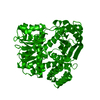
| Title | GLUTAMATE DEHYDROGENASE | ||||||
 Components Components | GLUTAMATE DEHYDROGENASE | ||||||
 Keywords Keywords | OXIDOREDUCTASE | ||||||
| Function / homology |  Function and homology information Function and homology information: / glutamate dehydrogenase (NAD+) / L-glutamate dehydrogenase (NADP+) activity / L-glutamate dehydrogenase (NAD+) activity / : / amino acid metabolic process / cytosol Similarity search - Function | ||||||
| Biological species |  Clostridium symbiosum (bacteria) Clostridium symbiosum (bacteria) | ||||||
| Method |  X-RAY DIFFRACTION / SYNCHROTRON / MIR / Resolution: 1.9 Å X-RAY DIFFRACTION / SYNCHROTRON / MIR / Resolution: 1.9 Å | ||||||
 Authors Authors | Stillman, T.J. / Baker, P.J. / Britton, K.L. / Rice, D.W. | ||||||
 Citation Citation | Journal: J.Mol.Biol. / Year: 1993 Title: Conformational flexibility in glutamate dehydrogenase. Role of water in substrate recognition and catalysis. Authors: Stillman, T.J. / Baker, P.J. / Britton, K.L. / Rice, D.W. #1: Journal: Biochemistry / Year: 1997Title: Determinants of Substrate Specificity in the Superfamily of Amino Acid Dehydrogenases Authors: Baker, P.J. / Waugh, M.L. / Wang, X.G. / Stillman, T.J. / Turnbull, A.P. / Engel, P.C. / Rice, D.W. #2: Journal: Structure / Year: 1995Title: The Structure of Pyrococcus Furiosus Glutamate Dehydrogenase Reveals a Key Role for Ion-Pair Networks in Maintaining Enzyme Stability at Extreme Temperatures Authors: Yip, K.S. / Stillman, T.J. / Britton, K.L. / Artymiuk, P.J. / Baker, P.J. / Sedelnikova, S.E. / Engel, P.C. / Pasquo, A. / Chiaraluce, R. / Consalvi, V. / Scandurra, R. / Rice, D.W. #3: Journal: J.Mol.Biol. / Year: 1993Title: Erratum. Structural Consequences of Sequence Patterns in the Fingerprint Region of the Nucleotide Binding Fold. Implications for Nucleotide Specificity Authors: Baker, P.J. / Britton, K.L. / Rice, D.W. / Rob, A. / Stillman, T.J. #4: Journal: J.Mol.Biol. / Year: 1992Title: Structural Consequences of Sequence Patterns in the Fingerprint Region of the Nucleotide Binding Fold. Implications for Nucleotide Specificity Authors: Baker, P.J. / Britton, K.L. / Rice, D.W. / Rob, A. / Stillman, T.J. #5: Journal: Proteins / Year: 1992Title: Subunit Assembly and Active Site Location in the Structure of Glutamate Dehydrogenase Authors: Baker, P.J. / Britton, K.L. / Engel, P.C. / Farrants, G.W. / Lilley, K.S. / Rice, D.W. / Stillman, T.J. #6: Journal: Eur.J.Biochem. / Year: 1992Title: Structural Relationship between the Hexameric and Tetrameric Family of Glutamate Dehydrogenases Authors: Britton, K.L. / Baker, P.J. / Rice, D.W. / Stillman, T.J. #7: Journal: J.Mol.Biol. / Year: 1985Title: Crystallization of an Nad+-Dependent Glutamate Dehydrogenase from Clostridium Symbiosum Authors: Rice, D.W. / Hornby, D.P. / Engel, P.C. | ||||||
| History |
|
- Structure visualization
| Structure viewer | Molecule: MolmilJmol/JSmol |
|---|
- Downloads & links
-Download
| PDBx/mmCIF format | 1bgv.cif.gz | 102.5 KB | Display | PDBx/mmCIF format |
|---|---|---|---|---|
| PDB format | pdb1bgv.ent.gz | 78.5 KB | Display | PDB format |
| PDBx/mmJSON format | 1bgv.json.gz | Tree view | PDBx/mmJSON format | |
| Others |  Other downloads Other downloads |
-Validation report
| Arichive directory | https://data.pdbj.org/pub/pdb/validation_reports/bg/1bgvftp://data.pdbj.org/pub/pdb/validation_reports/bg/1bgv | HTTPS FTP |
|---|
-Related structure data
| Similar structure data |
|---|
-Links
 PDBj
PDBj

- Assembly
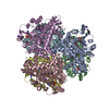
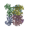
| Deposited unit | 
| ||||||||
|---|---|---|---|---|---|---|---|---|---|
| 1 | x 6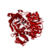
| ||||||||
| Unit cell |
|
-Components
| #1: Protein | Mass: 49215.648 Da / Num. of mol.: 1 / Source method: isolated from a natural source Details: THE RESIDUE LABELLED B 501, A GLUTAMATE, IS THE ENZYME'S SUBSTRATE WHICH IS BOUND WITHIN THE ACTIVE SITE. IT IS THOUGHT TO AN ONLY PARTIALLY OCCUPIED SITE AND SO ITS OCCUPANCY WAS SET TO 0.53. Source: (natural) Clostridium symbiosum (bacteria) / References: UniProt: P24295, glutamate dehydrogenase (NAD+) |
|---|---|
| #2: Chemical | ChemComp-GLU /   Type: L-peptide linking / Mass: 147.129 Da / Num. of mol.: 1 / Source method: obtained synthetically / Formula: C5H9NO4 Type: L-peptide linking / Mass: 147.129 Da / Num. of mol.: 1 / Source method: obtained synthetically / Formula: C5H9NO4 |
| #3: Water | ChemComp-HOH /  Mass: 18.015 Da / Num. of mol.: 217 / Source method: isolated from a natural source / Formula: H2O Mass: 18.015 Da / Num. of mol.: 217 / Source method: isolated from a natural source / Formula: H2O |
-Experimental details
-Experiment
| Experiment | Method: X-RAY DIFFRACTION / Number of used crystals: 3 |
|---|
- Sample preparation
| Crystal | Density Matthews: 2.6 Å3/Da / Density % sol: 54 % |
|---|---|
| Crystal grow | pH: 7 / Details: pH 7.0 |
-Data collection
| Diffraction | Mean temperature: 293 K |
|---|---|
| Diffraction source | Source: SYNCHROTRON / Site: SRS  / Beamline: PX9.5 / Wavelength: 0.88 / Beamline: PX9.5 / Wavelength: 0.88 |
| Detector | Detector: FILM / Date: Oct 7, 1991 |
| Radiation | Monochromatic (M) / Laue (L): M / Scattering type: x-ray |
| Radiation wavelength | Wavelength: 0.88 Å / Relative weight: 1 |
| Reflection | Resolution: 1.9→40 Å / Num. obs: 42325 / % possible obs: 99 % / Observed criterion σ(I): 0 / Redundancy: 3.36 % / Rmerge(I) obs: 0.07 |
- Processing
| Software |
| ||||||||||||||||||||||||||||||||||||||||||||||||||
|---|---|---|---|---|---|---|---|---|---|---|---|---|---|---|---|---|---|---|---|---|---|---|---|---|---|---|---|---|---|---|---|---|---|---|---|---|---|---|---|---|---|---|---|---|---|---|---|---|---|---|---|
| Refinement | Method to determine structure: MIR / Resolution: 1.9→10 Å / Isotropic thermal model: TNT / σ(F): 0 / Stereochemistry target values: ENGH AND HUBER Details: THE RESIDUE LABELLED B 501, A GLUTAMATE, IS THE ENZYME'S SUBSTRATE WHICH IS BOUND WITHIN THE ACTIVE SITE. IT IS THOUGHT TO AN ONLY PARTIALLY OCCUPIED SITE AND SO ITS OCCUPANCY WAS SET TO 0.53.
| ||||||||||||||||||||||||||||||||||||||||||||||||||
| Solvent computation | Solvent model: MOEWS AND KRETSINGER / Bsol: 248 Å2 / ksol: 0.85 e/Å3 | ||||||||||||||||||||||||||||||||||||||||||||||||||
| Refinement step | Cycle: LAST / Resolution: 1.9→10 Å
| ||||||||||||||||||||||||||||||||||||||||||||||||||
| Refine LS restraints |
|
