oxidoreductase activity, acting on the CH-NH2 group of donors, NAD or NADP as acceptor / amino acid metabolic process / nucleotide binding Similarity search - Function
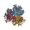
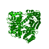
Glutamate Dehydrogenase, chain A, domain 3 / Glutamate Dehydrogenase; Chain A, domain 3 / Glutamate dehydrogenase / Leucine Dehydrogenase, chain A, domain 1 / NAD(P) binding domain of glutamate dehydrogenase / Leu/Phe/Val dehydrogenases active site / Glu / Leu / Phe / Val dehydrogenases active site. / Glutamate/phenylalanine/leucine/valine dehydrogenase / Glutamate/phenylalanine/leucine/valine dehydrogenase, dimerisation domain / Glu/Leu/Phe/Val dehydrogenase, dimerisation domain ...Glutamate Dehydrogenase, chain A, domain 3 / Glutamate Dehydrogenase; Chain A, domain 3 / Glutamate dehydrogenase / Leucine Dehydrogenase, chain A, domain 1 / NAD(P) binding domain of glutamate dehydrogenase / Leu/Phe/Val dehydrogenases active site / Glu / Leu / Phe / Val dehydrogenases active site. / Glutamate/phenylalanine/leucine/valine dehydrogenase / Glutamate/phenylalanine/leucine/valine dehydrogenase, dimerisation domain / Glu/Leu/Phe/Val dehydrogenase, dimerisation domain / Glutamate/Leucine/Phenylalanine/Valine dehydrogenase / Glutamate/phenylalanine/leucine/valine dehydrogenase, C-terminal / Glutamate/Leucine/Phenylalanine/Valine dehydrogenase / NAD(P)-binding Rossmann-like Domain / NAD(P)-binding domain superfamily / Rossmann fold / Orthogonal Bundle / 3-Layer(aba) Sandwich / Mainly Alpha / Alpha Beta Similarity search - Domain/homology
 Movie
Movie Controller

 Yorodumi
 Open data
 Basic information
 Components
 Keywords
 Function and homology information
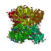
 Corynebacterium glutamicum (bacteria)
 X-RAY DIFFRACTION /
  Authors
 Japan, 1items
  Citation
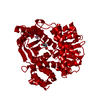
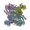
 Structure visualization
 Downloads & links
 Other downloads
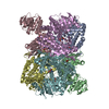

 PDBj
PDBj

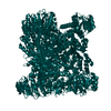
 Assembly
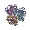
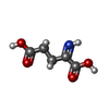


 Mass: 145.113 Da / Num. of mol.: 4 / Source method: obtained synthetically / Formula: C5H7NO4
 Mass: 745.421 Da / Num. of mol.: 6 / Source method: obtained synthetically / Formula: C21H30N7O17P3
 Mass: 39.098 Da / Num. of mol.: 14 / Source method: obtained synthetically / Formula: K
 Mass: 192.124 Da / Num. of mol.: 1 / Source method: obtained synthetically / Formula: C6H8O7
 Sample preparation
 Processing