Mass: 18.015 Da / Num. of mol.: 527 / Source method: isolated from a natural source / Formula: H2O
-
Details
Nonpolymer details
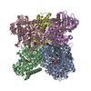
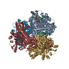
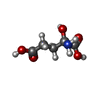
The author states that in this structure, the alpha-imino group of alpha-iminoglutarate is ...The author states that in this structure, the alpha-imino group of alpha-iminoglutarate is protonated. As this ligand is the reaction intermediate and the alpha-imino group forms an ionic interaction with the negatively charged carboxylate group of an active site asparate, it is highly possible that this group of the ligand is in the protonated form. The existing ligand (2IT) represents the neutral alpha-amino group, therefore does not satisfy the correct ionization state of the ligand present in this structure.
-
Experimental details
-
Experiment
Experiment
Method: X-RAY DIFFRACTION / Number of used crystals: 1
-
Sample preparation
Crystal grow
Temperature: 295 K / Method: vapor diffusion, sitting drop / pH: 6.5 Details: 40% v/v PEG 300, 0.1M sodium cacodylate, pH 6.5, 0.2 M calcium acetate hydrate
 Movie
Movie Controller

 Yorodumi
 Open data
 Basic information
 Components
 Keywords
 Function and homology information
 X-RAY DIFFRACTION /
  Authors
 India, 1items
  Citation
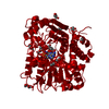
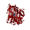
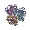
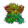
 Structure visualization
 Downloads & links
 Other downloads
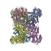


 PDBj
PDBj Assembly
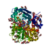


 Mass: 743.405 Da / Num. of mol.: 1 / Source method: obtained synthetically / Formula: C21H28N7O17P3
 Mass: 163.129 Da / Num. of mol.: 1 / Source method: obtained synthetically / Formula: C5H9NO5
 Mass: 145.113 Da / Num. of mol.: 1 / Source method: obtained synthetically / Formula: C5H7NO4
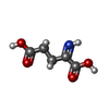
 Mass: 92.094 Da / Num. of mol.: 49 / Source method: obtained synthetically / Formula: C3H8O3
 Mass: 106.120 Da / Num. of mol.: 1 / Source method: obtained synthetically / Formula: C4H10O3
 Sample preparation
 / Beamline: BM14 / Wavelength: 0.9537 Å
 Processing