| Entry | Database: PDB / ID: 5xvx
|
|---|
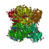
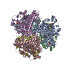
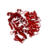
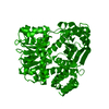
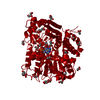
| Title | Crystal Structure of Aspergillus niger Glutamate Dehydrogenase Complexed With Alpha-ketoglutarate and NADPH |
|---|
 Components Components | Glutamate dehydrogenase |
|---|
 Keywords Keywords | OXIDOREDUCTASE / Aspergillus / Glutamate / Dehydrogenase / 2-Oxoglutarate / Allostery / NADPH / Coenzyme / Reaction mechanism |
|---|
| Function / homology |  Function and homology information Function and homology information
: / Glutamate dehydrogenase / NAD(P) binding domain of glutamate dehydrogenase / Leu/Phe/Val dehydrogenases active site / Glu / Leu / Phe / Val dehydrogenases active site. / Glutamate/phenylalanine/leucine/valine dehydrogenase / Glutamate/phenylalanine/leucine/valine dehydrogenase, dimerisation domain / Glu/Leu/Phe/Val dehydrogenase, dimerisation domain / Glutamate/Leucine/Phenylalanine/Valine dehydrogenase / Glutamate/phenylalanine/leucine/valine dehydrogenase, C-terminal ...: / Glutamate dehydrogenase / NAD(P) binding domain of glutamate dehydrogenase / Leu/Phe/Val dehydrogenases active site / Glu / Leu / Phe / Val dehydrogenases active site. / Glutamate/phenylalanine/leucine/valine dehydrogenase / Glutamate/phenylalanine/leucine/valine dehydrogenase, dimerisation domain / Glu/Leu/Phe/Val dehydrogenase, dimerisation domain / Glutamate/Leucine/Phenylalanine/Valine dehydrogenase / Glutamate/phenylalanine/leucine/valine dehydrogenase, C-terminal / Glutamate/Leucine/Phenylalanine/Valine dehydrogenase / Aminoacid dehydrogenase-like, N-terminal domain superfamily / NAD(P)-binding Rossmann-like Domain / NAD(P)-binding domain superfamily / Rossmann fold / 3-Layer(aba) Sandwich / Alpha BetaSimilarity search - Domain/homology |
|---|
| Biological species |   Aspergillus niger (mold) Aspergillus niger (mold) |
|---|
| Method |  X-RAY DIFFRACTION / SYNCHROTRON / MOLECULAR REPLACEMENT / Resolution: 1.8 Å X-RAY DIFFRACTION / SYNCHROTRON / MOLECULAR REPLACEMENT / Resolution: 1.8 Å |
|---|
 Authors Authors | Prakash, P. / Punekar, N.S. / Bhaumik, P. |
|---|
| Funding support |  India, 1items India, 1items | Organization | Grant number | Country |
|---|
| Department of Biotechnology | Ramalingaswami Re-entry Fellowship | India |
|
|---|
 Citation Citation | Journal: J. Biol. Chem. / Year: 2018
Title: Structural basis for the catalytic mechanism and alpha-ketoglutarate cooperativity of glutamate dehydrogenase.
Authors: Prakash, P. / Punekar, N.S. / Bhaumik, P. |
|---|
| History | | Deposition | Jun 28, 2017 | Deposition site: PDBJ / Processing site: PDBJ |
|---|
| Revision 1.0 | Mar 21, 2018 | Provider: repository / Type: Initial release |
|---|
| Revision 1.1 | Mar 28, 2018 | Group: Database references / Category: citation
Item: _citation.journal_abbrev / _citation.pdbx_database_id_PubMed / _citation.title |
|---|
| Revision 1.2 | May 9, 2018 | Group: Data collection / Database references / Category: citation
Item: _citation.journal_volume / _citation.page_first / _citation.page_last |
|---|
| Revision 1.3 | Mar 27, 2024 | Group: Data collection / Database references / Category: chem_comp_atom / chem_comp_bond / database_2
Item: _database_2.pdbx_DOI / _database_2.pdbx_database_accession |
|---|
|
|---|
 Movie
Movie Controller

 Yorodumi
 Open data
 Basic information
 Structure visualization
 Downloads & links
 Other downloads
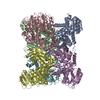


 PDBj
PDBj


 Assembly
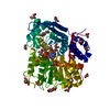


 Mass: 745.421 Da / Num. of mol.: 1 / Source method: obtained synthetically / Formula: C21H30N7O17P3
 Mass: 146.098 Da / Num. of mol.: 1 / Source method: obtained synthetically / Formula: C5H6O5
 Mass: 92.094 Da / Num. of mol.: 23 / Source method: obtained synthetically / Formula: C3H8O3
 Mass: 106.120 Da / Num. of mol.: 2 / Source method: obtained synthetically / Formula: C4H10O3
 Sample preparation
 / Beamline: BM14 / Wavelength: 0.9763 Å
 Processing