 Movie
Movie Controller

[English] 日本語
 Yorodumi
- PDB-5ijz: Crystal structure of glutamate dehydrogenase(GDH) from Corynebact... -
+ Open data

- Basic information
| Entry | Database: PDB / ID: 5ijz | ||||||
|---|---|---|---|---|---|---|---|
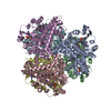
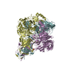
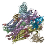
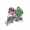
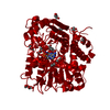
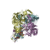
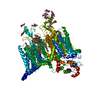
| Title | Crystal structure of glutamate dehydrogenase(GDH) from Corynebacterium glutamicum | ||||||
 Components Components | NADP-specific glutamate dehydrogenase | ||||||
 Keywords Keywords | OXIDOREDUCTASE / glutamate dehydrogenase | ||||||
| Function / homology |  Function and homology information Function and homology informationglutamate dehydrogenase (NADP+) / L-glutamate dehydrogenase (NADP+) activity / : / cytoplasm / cytosol Similarity search - Function | ||||||
| Biological species |  Corynebacterium glutamicum (bacteria) Corynebacterium glutamicum (bacteria) | ||||||
| Method |  X-RAY DIFFRACTION / SYNCHROTRON / MOLECULAR REPLACEMENT / Resolution: 2.29 Å X-RAY DIFFRACTION / SYNCHROTRON / MOLECULAR REPLACEMENT / Resolution: 2.29 Å | ||||||
 Authors Authors | Son, H.-F. / Kim, K.-J. | ||||||
 Citation Citation | Journal: BIOCHEM.BIOPHYS.RES.COMMUN. / Year: 2015 Title: Structural insights into domain movement and cofactor specificity of glutamate dehydrogenase from Corynebacterium glutamicum Authors: Son, H.-F. / Kim, K.-J. | ||||||
| History |
|
- Structure visualization
| Structure viewer | Molecule: MolmilJmol/JSmol |
|---|
- Downloads & links
-Download
| PDBx/mmCIF format | 5ijz.cif.gz | 1005.5 KB | Display | PDBx/mmCIF format |
|---|---|---|---|---|
| PDB format | pdb5ijz.ent.gz | 840.1 KB | Display | PDB format |
| PDBx/mmJSON format | 5ijz.json.gz | Tree view | PDBx/mmJSON format | |
| Others |  Other downloads Other downloads |
-Validation report
| Arichive directory | https://data.pdbj.org/pub/pdb/validation_reports/ij/5ijzftp://data.pdbj.org/pub/pdb/validation_reports/ij/5ijz | HTTPS FTP |
|---|
-Related structure data
| Related structure data |  1bgvS S: Starting model for refinement |
|---|---|
| Similar structure data |

-Links
 PDBj
PDBj

- Assembly
| Deposited unit | 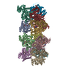
| ||||||||
|---|---|---|---|---|---|---|---|---|---|
| 1 | 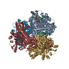
| ||||||||
| 2 | 
| ||||||||
| Unit cell |
|
-Components
| #1: Protein | Mass: 49046.102 Da / Num. of mol.: 12 / Fragment: Substrate-binding domain (UNP RESIDUES 1-420) Source method: isolated from a genetically manipulated source Source: (gene. exp.) Corynebacterium glutamicum (strain ATCC 13032 / DSM 20300 / JCM 1318 / LMG 3730 / NCIMB 10025) (bacteria)Strain: ATCC 13032 / DSM 20300 / JCM 1318 / LMG 3730 / NCIMB 10025 Gene: gdh, Cgl2079, cg2280 / Plasmid: pProEX-HTa / Production host: References: UniProt: P31026, glutamate dehydrogenase (NADP+) #2: Chemical | ChemComp-NAP /   Mass: 743.405 Da / Num. of mol.: 9 / Source method: obtained synthetically / Formula: C21H28N7O17P3 Mass: 743.405 Da / Num. of mol.: 9 / Source method: obtained synthetically / Formula: C21H28N7O17P3#3: Chemical | ChemComp-AKG /   Mass: 146.098 Da / Num. of mol.: 9 / Source method: isolated from a natural source / Formula: C5H6O5 Mass: 146.098 Da / Num. of mol.: 9 / Source method: isolated from a natural source / Formula: C5H6O5#4: Water | ChemComp-HOH / |  Mass: 18.015 Da / Num. of mol.: 1629 / Source method: isolated from a natural source / Formula: H2O Mass: 18.015 Da / Num. of mol.: 1629 / Source method: isolated from a natural source / Formula: H2O |
|---|
-Experimental details
-Experiment
| Experiment | Method: X-RAY DIFFRACTION / Number of used crystals: 1 |
|---|
- Sample preparation
| Crystal | Density Matthews: 2.42 Å3/Da / Density % sol: 49.08 % |
|---|---|
| Crystal grow | Temperature: 293 K / Method: vapor diffusion, hanging drop / pH: 5 / Details: PEG 3350, Tacsimate |
-Data collection
| Diffraction | Mean temperature: 100 K |
|---|---|
| Diffraction source | Source: SYNCHROTRON / Site: PAL/PLS  / Beamline: 7A (6B, 6C1) / Wavelength: 0.9793 Å / Beamline: 7A (6B, 6C1) / Wavelength: 0.9793 Å |
| Detector | Type: ADSC QUANTUM 270 / Detector: CCD / Date: Dec 23, 2014 |
| Radiation | Monochromator: Double Crystal Monochromator / Protocol: SINGLE WAVELENGTH / Monochromatic (M) / Laue (L): M / Scattering type: x-ray |
| Radiation wavelength | Wavelength: 0.9793 Å / Relative weight: 1 |
| Reflection | Resolution: 2.29→178.51 Å / Num. obs: 249267 / % possible obs: 98.6 % / Redundancy: 5.6 % / Net I/σ(I): 21.14 |
| Reflection shell | Resolution: 2.3→2.34 Å |
- Processing
| Software |
| |||||||||||||||||||||||||||||||||||||||||||||||||||||||||||||||||||||||||||
|---|---|---|---|---|---|---|---|---|---|---|---|---|---|---|---|---|---|---|---|---|---|---|---|---|---|---|---|---|---|---|---|---|---|---|---|---|---|---|---|---|---|---|---|---|---|---|---|---|---|---|---|---|---|---|---|---|---|---|---|---|---|---|---|---|---|---|---|---|---|---|---|---|---|---|---|---|
| Refinement | Method to determine structure: MOLECULAR REPLACEMENT Starting model: 1BGV Resolution: 2.29→34.366 Å / Cor.coef. Fo:Fc: 0.947 / Cor.coef. Fo:Fc free: 0.906 / SU B: 6.377 / SU ML: 0.156 / Cross valid method: THROUGHOUT / σ(F): 0 / ESU R: 0.315 / ESU R Free: 0.223 / Stereochemistry target values: MAXIMUM LIKELIHOOD Details: HYDROGENS HAVE BEEN ADDED IN THE RIDING POSITIONS U VALUES
| |||||||||||||||||||||||||||||||||||||||||||||||||||||||||||||||||||||||||||
| Solvent computation | Ion probe radii: 0.8 Å / Shrinkage radii: 0.8 Å / VDW probe radii: 1.2 Å / Solvent model: MASK | |||||||||||||||||||||||||||||||||||||||||||||||||||||||||||||||||||||||||||
| Displacement parameters | Biso max: 175.34 Å2 / Biso mean: 25.987 Å2 / Biso min: 5.41 Å2
| |||||||||||||||||||||||||||||||||||||||||||||||||||||||||||||||||||||||||||
| Refinement step | Cycle: final / Resolution: 2.29→34.366 Å
| |||||||||||||||||||||||||||||||||||||||||||||||||||||||||||||||||||||||||||
| Refine LS restraints |
| |||||||||||||||||||||||||||||||||||||||||||||||||||||||||||||||||||||||||||
| LS refinement shell | Resolution: 2.293→2.353 Å / Total num. of bins used: 20
|
