 Movie
Movie Controller

[English] 日本語
 Yorodumi
- PDB-6hjy: X-ray structure of a pentameric ligand gated ion channel from Erw... -
+ Open data

- Basic information
| Entry | Database: PDB / ID: 6hjy | ||||||
|---|---|---|---|---|---|---|---|
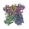
| Title | X-ray structure of a pentameric ligand gated ion channel from Erwinia chrysanthemi (ELIC) Delta8 truncation mutant in complex with nanobody 72 | ||||||
 Components Components |
| ||||||
 Keywords Keywords | MEMBRANE PROTEIN / ion channel / pentameric ligand-gated ion channel / cya-loop receptor | ||||||
| Function / homology |  Function and homology information Function and homology informationextracellular ligand-gated monoatomic ion channel activity / transmembrane signaling receptor activity / identical protein binding / plasma membrane Similarity search - Function | ||||||
| Biological species |  Dickeya chrysanthemi (bacteria) Dickeya chrysanthemi (bacteria) | ||||||
| Method |  X-RAY DIFFRACTION / SYNCHROTRON / MOLECULAR REPLACEMENT / Resolution: 2.78 Å X-RAY DIFFRACTION / SYNCHROTRON / MOLECULAR REPLACEMENT / Resolution: 2.78 Å | ||||||
 Authors Authors | Spurny, R. / Govaerts, C. / Evans, G.L. / Pardon, E. / Steyaert, J. / Ulens, C. | ||||||
 Citation Citation | Journal: Nat.Chem.Biol. / Year: 2019 Title: A lipid site shapes the agonist response of a pentameric ligand-gated ion channel. Authors: Henault, C.M. / Govaerts, C. / Spurny, R. / Brams, M. / Estrada-Mondragon, A. / Lynch, J. / Bertrand, D. / Pardon, E. / Evans, G.L. / Woods, K. / Elberson, B.W. / Cuello, L.G. / Brannigan, G. ...Authors: Henault, C.M. / Govaerts, C. / Spurny, R. / Brams, M. / Estrada-Mondragon, A. / Lynch, J. / Bertrand, D. / Pardon, E. / Evans, G.L. / Woods, K. / Elberson, B.W. / Cuello, L.G. / Brannigan, G. / Nury, H. / Steyaert, J. / Baenziger, J.E. / Ulens, C. | ||||||
| History |
|
- Structure visualization
| Structure viewer | Molecule: MolmilJmol/JSmol |
|---|
- Downloads & links
-Download
| PDBx/mmCIF format | 6hjy.cif.gz | 402.2 KB | Display | PDBx/mmCIF format |
|---|---|---|---|---|
| PDB format | pdb6hjy.ent.gz | 325.6 KB | Display | PDB format |
| PDBx/mmJSON format | 6hjy.json.gz | Tree view | PDBx/mmJSON format | |
| Others |  Other downloads Other downloads |
-Validation report
| Arichive directory | https://data.pdbj.org/pub/pdb/validation_reports/hj/6hjyftp://data.pdbj.org/pub/pdb/validation_reports/hj/6hjy | HTTPS FTP |
|---|
-Related structure data
| Related structure data |  6hjxSC  6hk0C S: Starting model for refinement C: citing same article ( |
|---|---|
| Similar structure data |

-Links
 PDBj
PDBj


- Assembly
| Deposited unit | 
| ||||||||
|---|---|---|---|---|---|---|---|---|---|
| 1 |
| ||||||||
| Unit cell |
|
-Components
-Cys-loop ligand-gated ion ... , 4 types, 5 molecules ABCDE
| #1: Protein | Mass: 32102.461 Da / Num. of mol.: 1 Source method: isolated from a genetically manipulated source Source: (gene. exp.) Dickeya chrysanthemi (bacteria) / Production host: | ||||
|---|---|---|---|---|---|
| #2: Protein | Mass: 32413.795 Da / Num. of mol.: 2 Source method: isolated from a genetically manipulated source Source: (gene. exp.) Dickeya chrysanthemi (bacteria) / Production host: #3: Protein | | Mass: 32031.383 Da / Num. of mol.: 1 Source method: isolated from a genetically manipulated source Source: (gene. exp.) Dickeya chrysanthemi (bacteria) / Production host: #4: Protein | | Mass: 32217.547 Da / Num. of mol.: 1 Source method: isolated from a genetically manipulated source Source: (gene. exp.) Dickeya chrysanthemi (bacteria) / Production host: |
-Antibody , 2 types, 5 molecules FJGHI
| #5: Antibody | Mass: 13367.960 Da / Num. of mol.: 2 Source method: isolated from a genetically manipulated source Source: (gene. exp.) #6: Antibody | Mass: 13239.831 Da / Num. of mol.: 3 Source method: isolated from a genetically manipulated source Source: (gene. exp.) |
|---|
-Details
| Has protein modification | Y |
|---|
-Experimental details
-Experiment
| Experiment | Method: X-RAY DIFFRACTION / Number of used crystals: 1 |
|---|
- Sample preparation
| Crystal | Density Matthews: 3.23 Å3/Da / Density % sol: 61.87 % |
|---|---|
| Crystal grow | Temperature: 293 K / Method: vapor diffusion, sitting drop Details: 300 mM ammonium formate, 50 mM TRIS pH 9.0 and 33% PEG monomethylether 550 |
-Data collection
| Diffraction | Mean temperature: 100 K / Ambient temp details: 100 / Serial crystal experiment: N |
|---|---|
| Diffraction source | Source: SYNCHROTRON / Site: ESRF  / Beamline: ID30B / Wavelength: 0.9754 Å / Beamline: ID30B / Wavelength: 0.9754 Å |
| Detector | Type: DECTRIS PILATUS 300K / Detector: PIXEL / Date: Oct 24, 2017 |
| Radiation | Protocol: SINGLE WAVELENGTH / Monochromatic (M) / Laue (L): M / Scattering type: x-ray |
| Radiation wavelength | Wavelength: 0.9754 Å / Relative weight: 1 |
| Reflection | Resolution: 2.78→49.49 Å / Num. obs: 71824 / % possible obs: 99.3 % / Redundancy: 3.3 % / Biso Wilson estimate: 88.12 Å2 / CC1/2: 0.995 / Rmerge(I) obs: 0.098 / Rpim(I) all: 0.094 / Rrim(I) all: 0.136 / Net I/av σ(I): 6.6 / Net I/σ(I): 6.6 |
| Reflection shell | Resolution: 2.78→2.85 Å / Rmerge(I) obs: 1.442 / Mean I/σ(I) obs: 0.6 / Num. unique obs: 4318 / CC1/2: 0.281 / Rpim(I) all: 1.379 / Rrim(I) all: 1.998 |
- Processing
| Software |
| ||||||||||||||||||||||||||||||||||||||||||||||||||||||||||||||||||||||||||||||||||||||||||||||||||||||||||||
|---|---|---|---|---|---|---|---|---|---|---|---|---|---|---|---|---|---|---|---|---|---|---|---|---|---|---|---|---|---|---|---|---|---|---|---|---|---|---|---|---|---|---|---|---|---|---|---|---|---|---|---|---|---|---|---|---|---|---|---|---|---|---|---|---|---|---|---|---|---|---|---|---|---|---|---|---|---|---|---|---|---|---|---|---|---|---|---|---|---|---|---|---|---|---|---|---|---|---|---|---|---|---|---|---|---|---|---|---|---|
| Refinement | Method to determine structure: MOLECULAR REPLACEMENT Starting model: 6HJX Resolution: 2.78→49.49 Å / Cor.coef. Fo:Fc: 0.901 / Cor.coef. Fo:Fc free: 0.912 / SU R Cruickshank DPI: 0.919 / Cross valid method: THROUGHOUT / σ(F): 0 / SU R Blow DPI: 0.712 / SU Rfree Blow DPI: 0.292 / SU Rfree Cruickshank DPI: 0.307
| ||||||||||||||||||||||||||||||||||||||||||||||||||||||||||||||||||||||||||||||||||||||||||||||||||||||||||||
| Displacement parameters | Biso max: 253.68 Å2 / Biso mean: 101.39 Å2 / Biso min: 45.07 Å2
| ||||||||||||||||||||||||||||||||||||||||||||||||||||||||||||||||||||||||||||||||||||||||||||||||||||||||||||
| Refine analyze | Luzzati coordinate error obs: 0.45 Å | ||||||||||||||||||||||||||||||||||||||||||||||||||||||||||||||||||||||||||||||||||||||||||||||||||||||||||||
| Refinement step | Cycle: final / Resolution: 2.78→49.49 Å
| ||||||||||||||||||||||||||||||||||||||||||||||||||||||||||||||||||||||||||||||||||||||||||||||||||||||||||||
| Refine LS restraints |
| ||||||||||||||||||||||||||||||||||||||||||||||||||||||||||||||||||||||||||||||||||||||||||||||||||||||||||||
| LS refinement shell | Resolution: 2.78→2.85 Å / Rfactor Rfree error: 0 / Total num. of bins used: 20
|
