 Movie
Movie Controller


+ Open data

- Basic information
| Entry | Database: PDB / ID: 1bvu | ||||||
|---|---|---|---|---|---|---|---|
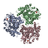
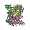
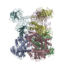
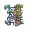
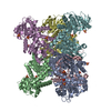
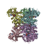
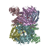
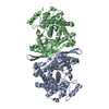
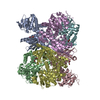
| Title | GLUTAMATE DEHYDROGENASE FROM THERMOCOCCUS LITORALIS | ||||||
 Components Components | PROTEIN (GLUTAMATE DEHYDROGENASE) | ||||||
 Keywords Keywords | OXIDOREDUCTASE / Thermal Stability | ||||||
| Function / homology |  Function and homology information Function and homology informationglutamate dehydrogenase [NAD(P)+] / L-glutamate dehydrogenase (NADP+) activity / L-glutamate dehydrogenase (NAD+) activity / L-glutamate catabolic process / cytoplasm Similarity search - Function | ||||||
| Biological species |   Thermococcus litoralis (archaea) Thermococcus litoralis (archaea) | ||||||
| Method |  X-RAY DIFFRACTION / SYNCHROTRON / MIR / Resolution: 2.5 Å X-RAY DIFFRACTION / SYNCHROTRON / MIR / Resolution: 2.5 Å | ||||||
 Authors Authors | Baker, P.J. / Britton, K.L. / Yip, K.S. / Stillman, T.J. / Rice, D.W. | ||||||
 Citation Citation | Journal: J.Mol.Biol. / Year: 1999 Title: Structure determination of the glutamate dehydrogenase from the hyperthermophile Thermococcus litoralis and its comparison with that from Pyrococcus furiosus Authors: Britton, K.L. / Yip, K.S. / Sedelnikova, S.E. / Stillman, T.J. / Adams, M.W. / Ma, K. / Maeder, D.L. / Robb, F.T. / Tolliday, N. / Vetriani, C. / Rice, D.W. / Baker, P.J. #1: Journal: Proc.Natl.Acad.Sci.USA / Year: 1998Title: Protein Thermostability Above 100C: A Key Role for Ionic Interactions Authors: Vetriani, C. / Maeder, D.L. / Tolliday, N. / Yip, K.S.-P. / Stillman, T.J. / Britton, K.L. / Rice, D.W. / Klump, H.H. / Robb, F.T. #2: Journal: Acta Crystallogr.,Sect.D / Year: 1996Title: Crystallisation of the Glutamate Dehydrogenase from the Hyperthermophilic Archaeon Thermococcus Litoralis Authors: Sedelnikova, S.E. / Yip, K.S.-P. / Stillman, T.J. / Ma, K. / Adams, M.W.W. / Robb, F.T. / Rice, D.W. | ||||||
| History |
|
- Structure visualization
| Structure viewer | Molecule: MolmilJmol/JSmol |
|---|
- Downloads & links
-Download
| PDBx/mmCIF format | 1bvu.cif.gz | 466.7 KB | Display | PDBx/mmCIF format |
|---|---|---|---|---|
| PDB format | pdb1bvu.ent.gz | 389.2 KB | Display | PDB format |
| PDBx/mmJSON format | 1bvu.json.gz | Tree view | PDBx/mmJSON format | |
| Others |  Other downloads Other downloads |
-Validation report
| Arichive directory | https://data.pdbj.org/pub/pdb/validation_reports/bv/1bvuftp://data.pdbj.org/pub/pdb/validation_reports/bv/1bvu | HTTPS FTP |
|---|
-Related structure data
| Similar structure data |
|---|

-Links
 PDBj
PDBj- Assembly
| Deposited unit | 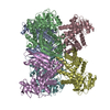
| ||||||||
|---|---|---|---|---|---|---|---|---|---|
| 1 |
| ||||||||
| Unit cell |
|
-Components
| #1: Protein | Mass: 46664.219 Da / Num. of mol.: 6 / Source method: isolated from a natural source / Source: (natural) Thermococcus litoralis (archaea)References: UniProt: Q56304, glutamate dehydrogenase [NAD(P)+] |
|---|
-Experimental details
-Experiment
| Experiment | Method: X-RAY DIFFRACTION |
|---|
- Sample preparation
| Crystal | Density Matthews: 2.88 Å3/Da / Density % sol: 57.33 % | |||||||||||||||||||||||||
|---|---|---|---|---|---|---|---|---|---|---|---|---|---|---|---|---|---|---|---|---|---|---|---|---|---|---|
| Crystal grow | pH: 8 Details: 35MG/ML PROTEIN IN 0.1M HEPES BUFFER PH 8.0 CONTAINING 1.7-1.8M AMMONIUM SULPHATE AND 1.5% W/V PEG 8000. | |||||||||||||||||||||||||
| Crystal grow | *PLUS Method: vapor diffusion, hanging drop | |||||||||||||||||||||||||
| Components of the solutions | *PLUS
|
-Data collection
| Diffraction | Mean temperature: 293 K |
|---|---|
| Diffraction source | Source: SYNCHROTRON / Site: SRS  / Beamline: PX9.5 / Wavelength: 0.92 / Beamline: PX9.5 / Wavelength: 0.92 |
| Detector | Type: MARRESEARCH / Detector: IMAGE PLATE |
| Radiation | Protocol: SINGLE WAVELENGTH / Monochromatic (M) / Laue (L): M / Scattering type: x-ray |
| Radiation wavelength | Wavelength: 0.92 Å / Relative weight: 1 |
| Reflection | Resolution: 2.5→20 Å / Num. obs: 101702 / % possible obs: 93 % / Observed criterion σ(I): 0 / Redundancy: 1.7 % / Rmerge(I) obs: 0.042 |
| Reflection | *PLUS Num. measured all: 173711 |
- Processing
| Software |
| ||||||||||||||||||||||||||||||
|---|---|---|---|---|---|---|---|---|---|---|---|---|---|---|---|---|---|---|---|---|---|---|---|---|---|---|---|---|---|---|---|
| Refinement | Method to determine structure: MIR / Resolution: 2.5→10 Å / σ(F): 0 / Stereochemistry target values: PROTGEO /
| ||||||||||||||||||||||||||||||
| Refinement step | Cycle: LAST / Resolution: 2.5→10 Å
| ||||||||||||||||||||||||||||||
| Refine LS restraints |
| ||||||||||||||||||||||||||||||
| Software | *PLUS Name: TNT / Classification: refinement | ||||||||||||||||||||||||||||||
| Refinement | *PLUS Rfactor Rwork: 0.192 | ||||||||||||||||||||||||||||||
| Solvent computation | *PLUS | ||||||||||||||||||||||||||||||
| Displacement parameters | *PLUS |
