 Movie
Movie Controller


+ Open data

- Basic information
| Entry | Database: PDB / ID: 1gtm | ||||||
|---|---|---|---|---|---|---|---|
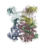
| Title | STRUCTURE OF GLUTAMATE DEHYDROGENASE | ||||||
 Components Components | GLUTAMATE DEHYDROGENASE | ||||||
 Keywords Keywords | OXIDOREDUCTASE / NAD / NADP | ||||||
| Function / homology |  Function and homology information Function and homology informationglutamate dehydrogenase [NAD(P)+] / L-glutamate dehydrogenase (NADP+) activity / L-glutamate dehydrogenase (NAD+) activity / L-glutamate catabolic process / cytoplasm Similarity search - Function | ||||||
| Biological species |   Pyrococcus furiosus (archaea) Pyrococcus furiosus (archaea) | ||||||
| Method |  X-RAY DIFFRACTION / SYNCHROTRON / Resolution: 2.2 Å X-RAY DIFFRACTION / SYNCHROTRON / Resolution: 2.2 Å | ||||||
 Authors Authors | Yip, K.S.P. / Stillman, T.J. / Britton, K.L. / Pasquo, A. / Rice, D.W. | ||||||
 Citation Citation | Journal: Structure / Year: 1995 Title: The structure of Pyrococcus furiosus glutamate dehydrogenase reveals a key role for ion-pair networks in maintaining enzyme stability at extreme temperatures. Authors: Yip, K.S. / Stillman, T.J. / Britton, K.L. / Artymiuk, P.J. / Baker, P.J. / Sedelnikova, S.E. / Engel, P.C. / Pasquo, A. / Chiaraluce, R. / Consalvi, V. / Scandurra, R. / Rice, D.W. #1: Journal: Eur.J.Biochem. / Year: 1995Title: Insights Into Thermal Stability from a Comparison of the Glutamate Dehydrogenases from Pyrococcus Furiosus and Thermococcus Litoralis Authors: Britton, K.L. / Baker, P.J. / Borges, K.M. / Engel, P.C. / Pasquo, A. / Rice, D.W. / Robb, F.T. / Scandurra, R. / Stillman, T.J. / Yip, K.S. #2: Journal: Acta Crystallogr.,Sect.D / Year: 1995Title: Crystallisation of the Nad(P)-Dependent Glutamate Dehydrogenase from the Hyperthermophile Pyrococcus Furiosus Authors: Yip, K.S.P. / Stillman, T.J. / Baker, P.J. / Britton, K.L. / Engel, P.C. / Sedelnikova, S.E. / Rice, D.W. / Pasquo, A. / Chiaraluce, R. / Consalvi, V. / Scandurra, R. | ||||||
| History |
|
- Structure visualization
| Structure viewer | Molecule: MolmilJmol/JSmol |
|---|
- Downloads & links
-Download
| PDBx/mmCIF format | 1gtm.cif.gz | 252.7 KB | Display | PDBx/mmCIF format |
|---|---|---|---|---|
| PDB format | pdb1gtm.ent.gz | 206.9 KB | Display | PDB format |
| PDBx/mmJSON format | 1gtm.json.gz | Tree view | PDBx/mmJSON format | |
| Others |  Other downloads Other downloads |
-Validation report
| Arichive directory | https://data.pdbj.org/pub/pdb/validation_reports/gt/1gtmftp://data.pdbj.org/pub/pdb/validation_reports/gt/1gtm | HTTPS FTP |
|---|
-Related structure data


-Links
 PDBj
PDBj- Assembly
| Deposited unit | 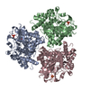
| ||||||||||||||||||||||||||||
|---|---|---|---|---|---|---|---|---|---|---|---|---|---|---|---|---|---|---|---|---|---|---|---|---|---|---|---|---|---|
| 1 | 
| ||||||||||||||||||||||||||||
| Unit cell |
| ||||||||||||||||||||||||||||
| Noncrystallographic symmetry (NCS) | NCS oper:
|
-Components
| #1: Protein | Mass: 46982.590 Da / Num. of mol.: 3 / Source method: isolated from a natural source / Source: (natural) Pyrococcus furiosus (archaea) / Strain: DSM 3638References: UniProt: P80319, glutamate dehydrogenase [NAD(P)+] #2: Chemical | ChemComp-SO4 /   Mass: 96.063 Da / Num. of mol.: 6 / Source method: obtained synthetically / Formula: SO4 Mass: 96.063 Da / Num. of mol.: 6 / Source method: obtained synthetically / Formula: SO4#3: Water | ChemComp-HOH / |  Mass: 18.015 Da / Num. of mol.: 318 / Source method: isolated from a natural source / Formula: H2O Mass: 18.015 Da / Num. of mol.: 318 / Source method: isolated from a natural source / Formula: H2O |
|---|
-Experimental details
-Experiment
| Experiment | Method: X-RAY DIFFRACTION |
|---|
- Sample preparation
| Crystal | Density Matthews: 4.28 Å3/Da / Density % sol: 67.4 % | ||||||||||||||||||||
|---|---|---|---|---|---|---|---|---|---|---|---|---|---|---|---|---|---|---|---|---|---|
| Crystal | *PLUS | ||||||||||||||||||||
| Crystal grow | *PLUS Temperature: 290 K / pH: 7.5 / Method: vapor diffusion, hanging dropDetails: Yip, K.S.P., (1995) Acta Crystallogr.,Sect.D, 51, 240. | ||||||||||||||||||||
| Components of the solutions | *PLUS
|
-Data collection
| Diffraction source | Source: SYNCHROTRON / Site: SRS  / Beamline: PX9.5 / Wavelength: 0.92 / Beamline: PX9.5 / Wavelength: 0.92 |
|---|---|
| Detector | Type: MARRESEARCH / Detector: IMAGE PLATE / Date: Sep 1, 1994 |
| Radiation | Monochromatic (M) / Laue (L): M / Scattering type: x-ray |
| Radiation wavelength | Wavelength: 0.92 Å / Relative weight: 1 |
| Reflection | Num. obs: 115181 / % possible obs: 93.2 % / Observed criterion σ(I): 0 / Redundancy: 2.6 % / Rmerge(I) obs: 0.043 |
| Reflection | *PLUS Highest resolution: 2.2 Å / Redundancy: 2.58 % |
- Processing
| Software |
| ||||||||||||||||||||||||||||||
|---|---|---|---|---|---|---|---|---|---|---|---|---|---|---|---|---|---|---|---|---|---|---|---|---|---|---|---|---|---|---|---|
| Refinement | Resolution: 2.2→10 Å
| ||||||||||||||||||||||||||||||
| Refinement step | Cycle: LAST / Resolution: 2.2→10 Å
| ||||||||||||||||||||||||||||||
| Refine LS restraints |
| ||||||||||||||||||||||||||||||
| Software | *PLUS Name: TNT / Classification: refinement | ||||||||||||||||||||||||||||||
| Refinement | *PLUS Num. reflection obs: 115153 / Rfactor obs: 0.17 | ||||||||||||||||||||||||||||||
| Solvent computation | *PLUS | ||||||||||||||||||||||||||||||
| Displacement parameters | *PLUS Biso mean: 38 Å2 | ||||||||||||||||||||||||||||||
| Refine LS restraints | *PLUS Type: t_angle_deg / Dev ideal: 2.85 |
