 Movie
Movie Controller


+ Open data

- Basic information
| Entry | Database: EMDB / ID: EMD-6495 | |||||||||
|---|---|---|---|---|---|---|---|---|---|---|
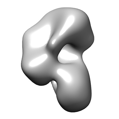
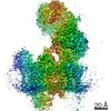
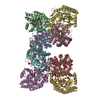
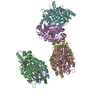
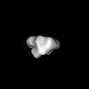
| Title | Electron microscopy of apo human XPC complex | |||||||||
 Map data Map data | EMAN2 reconstruction of apo human XPC complex | |||||||||
 Sample Sample |
| |||||||||
 Keywords Keywords | transcription / DNA repair / stem cells | |||||||||
| Function / homology |  Function and homology information Function and homology informationheteroduplex DNA loop binding / nucleotide-excision repair factor 2 complex / pyrimidine dimer repair by nucleotide-excision repair / XPC complex / 9+2 motile cilium / nucleotide-excision repair complex / photoreceptor connecting cilium / DNA damage sensor activity / regulation of proteasomal ubiquitin-dependent protein catabolic process / heterotrimeric G-protein binding ...heteroduplex DNA loop binding / nucleotide-excision repair factor 2 complex / pyrimidine dimer repair by nucleotide-excision repair / XPC complex / 9+2 motile cilium / nucleotide-excision repair complex / photoreceptor connecting cilium / DNA damage sensor activity / regulation of proteasomal ubiquitin-dependent protein catabolic process / heterotrimeric G-protein binding / transcription export complex 2 / histone H4K20 demethylase activity / nuclear pore nuclear basket / response to auditory stimulus / bubble DNA binding / Oxidoreductases; Acting on paired donors, with incorporation or reduction of molecular oxygen; With 2-oxoglutarate as one donor, and incorporation of one atom of oxygen into each donor / cellular response to interleukin-7 / response to UV-B / mitotic intra-S DNA damage checkpoint signaling / UV-damage excision repair / regulation of mitotic cell cycle phase transition / proteasome binding / centriole replication / embryonic organ development / glial cell projection / mRNA transport / polyubiquitin modification-dependent protein binding / mismatch repair / SUMOylation of DNA damage response and repair proteins / site of DNA damage / Loss of Nlp from mitotic centrosomes / Loss of proteins required for interphase microtubule organization from the centrosome / Recruitment of mitotic centrosome proteins and complexes / Recruitment of NuMA to mitotic centrosomes / Anchoring of the basal body to the plasma membrane / AURKA Activation by TPX2 / proteasome complex / regulation of cytokinesis / Josephin domain DUBs / nucleotide-excision repair / ubiquitin binding / centriole / N-glycan trimming in the ER and Calnexin/Calreticulin cycle / microtubule cytoskeleton organization / DNA Damage Recognition in GG-NER / G-protein beta/gamma-subunit complex binding / apical part of cell / Formation of Incision Complex in GG-NER / Regulation of PLK1 Activity at G2/M Transition / mitotic cell cycle / protein transport / single-stranded DNA binding / spermatogenesis / microtubule binding / damaged DNA binding / RNA polymerase II-specific DNA-binding transcription factor binding / proteasome-mediated ubiquitin-dependent protein catabolic process / transcription coactivator activity / ciliary basal body / RNA polymerase II cis-regulatory region sequence-specific DNA binding / response to xenobiotic stimulus / cell division / DNA repair / calcium ion binding / centrosome / positive regulation of DNA-templated transcription / chromatin / protein-containing complex binding / nucleolus / mitochondrion / nucleoplasm / nucleus / plasma membrane / cytoplasm / cytosol Similarity search - Function | |||||||||
| Biological species |  Homo sapiens (human) Homo sapiens (human) | |||||||||
| Method | single particle reconstruction / negative staining / Resolution: 25.0 Å | |||||||||
 Authors Authors | Zhang ET / He Y / Grob P / Fong YW / Nogales E / Tjian R | |||||||||
 Citation Citation | Journal: Proc Natl Acad Sci U S A / Year: 2015 Title: Architecture of the human XPC DNA repair and stem cell coactivator complex. Authors: Elisa T Zhang / Yuan He / Patricia Grob / Yick W Fong / Eva Nogales / Robert Tjian /  Abstract: The Xeroderma pigmentosum complementation group C (XPC) complex is a versatile factor involved in both nucleotide excision repair and transcriptional coactivation as a critical component of the ...The Xeroderma pigmentosum complementation group C (XPC) complex is a versatile factor involved in both nucleotide excision repair and transcriptional coactivation as a critical component of the NANOG, OCT4, and SOX2 pluripotency gene regulatory network. Here we present the structure of the human holo-XPC complex determined by single-particle electron microscopy to reveal a flexible, ear-shaped structure that undergoes localized loss of order upon DNA binding. We also determined the structure of the complete yeast homolog Rad4 holo-complex to find a similar overall architecture to the human complex, consistent with their shared DNA repair functions. Localized differences between these structures reflect an intriguing phylogenetic divergence in transcriptional capabilities that we present here. Having positioned the constituent subunits by tagging and deletion, we propose a model of key interaction interfaces that reveals the structural basis for this difference in functional conservation. Together, our findings establish a framework for understanding the structure-function relationships of the XPC complex in the interplay between transcription and DNA repair. | |||||||||
| History |
|
- Structure visualization
| Movie |
Movie viewer |
|---|---|
| Structure viewer | EM map: SurfViewMolmilJmol/JSmol |
| Supplemental images |
- Downloads & links
-EMDB archive
| Map data | emd_6495.map.gz | 6.4 MB | EMDB map data format | |
|---|---|---|---|---|
| Header (meta data) | emd-6495-v30.xmlemd-6495.xml | 13.6 KB 13.6 KB | Display Display | EMDB header |
| Images | emd_6495.tif | 755.8 KB | ||
| Archive directory |  http://ftp.pdbj.org/pub/emdb/structures/EMD-6495ftp://ftp.pdbj.org/pub/emdb/structures/EMD-6495 http://ftp.pdbj.org/pub/emdb/structures/EMD-6495ftp://ftp.pdbj.org/pub/emdb/structures/EMD-6495 | HTTPS FTP |
-Related structure data


-Links
| EMDB pages | EMDB (EBI/PDBe) / EMDataResource |
|---|---|
| Related items in Molecule of the Month |


-Map
| File | Download / File: emd_6495.map.gz / Format: CCP4 / Size: 7.8 MB / Type: IMAGE STORED AS FLOATING POINT NUMBER (4 BYTES) | ||||||||||||||||||||||||||||||||||||||||||||||||||||||||||||||||||||
|---|---|---|---|---|---|---|---|---|---|---|---|---|---|---|---|---|---|---|---|---|---|---|---|---|---|---|---|---|---|---|---|---|---|---|---|---|---|---|---|---|---|---|---|---|---|---|---|---|---|---|---|---|---|---|---|---|---|---|---|---|---|---|---|---|---|---|---|---|---|
| Annotation | EMAN2 reconstruction of apo human XPC complex | ||||||||||||||||||||||||||||||||||||||||||||||||||||||||||||||||||||
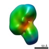
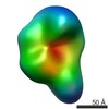
| Projections & slices | Image control
Images are generated by Spider. | ||||||||||||||||||||||||||||||||||||||||||||||||||||||||||||||||||||
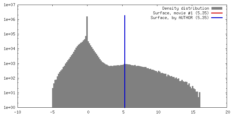
| Voxel size | X=Y=Z: 3.01 Å | ||||||||||||||||||||||||||||||||||||||||||||||||||||||||||||||||||||
| Density |
| ||||||||||||||||||||||||||||||||||||||||||||||||||||||||||||||||||||
| Symmetry | Space group: 1 | ||||||||||||||||||||||||||||||||||||||||||||||||||||||||||||||||||||
| Details | EMDB XML:
CCP4 map header:
| ||||||||||||||||||||||||||||||||||||||||||||||||||||||||||||||||||||
 Z (Sec.)
 Y (Row.)
 X (Col.)


-Supplemental data
- Sample components
-Entire : Apo human XPC complex
| Entire | Name: Apo human XPC complex |
|---|---|
| Components |
|
-Supramolecule #1000: Apo human XPC complex
| Supramolecule | Name: Apo human XPC complex / type: sample / ID: 1000 Details: Monodisperse. Thawed from -80 degrees Celsius and placed on ice immediately prior to grid preparation. Oligomeric state: heterotrimer / Number unique components: 3 |
|---|---|
| Molecular weight | Experimental: 200 KDa / Theoretical: 200 KDa / Method: SDS-PAGE, size exclusion chromatography |
-Macromolecule #1: Xeroderma pigmentosum group C-complementing protein
| Macromolecule | Name: Xeroderma pigmentosum group C-complementing protein / type: protein_or_peptide / ID: 1 Name.synonym: XPC, p125, DNA repair protein complementing XP-C cells Details: contains N-terminal 6xHis and TEV cleavage site / Number of copies: 1 / Oligomeric state: monomer / Recombinant expression: Yes |
|---|---|
| Source (natural) | Organism: Homo sapiens (human) / synonym: human / Location in cell: nucleus, cytoplasm |
| Molecular weight | Experimental: 125 KDa / Theoretical: 125 KDa |
| Recombinant expression | Organism:   Spodoptera frugiperda (fall armyworm) / Recombinant cell: Sf9 Spodoptera frugiperda (fall armyworm) / Recombinant cell: Sf9 |
| Sequence | UniProtKB: DNA repair protein complementing XP-C cells / GO: XPC complex |
-Macromolecule #2: RAD23 homolog B
| Macromolecule | Name: RAD23 homolog B / type: protein_or_peptide / ID: 2 Name.synonym: RAD23B, HR23B, HHR23B, p58, XP-C repair-complementing complex 58 kDa protein Details: contains N-terminal 1xFLAG tag / Number of copies: 1 / Oligomeric state: monomer / Recombinant expression: Yes |
|---|---|
| Source (natural) | Organism: Homo sapiens (human) / synonym: human / Location in cell: nucleus, cytoplasm |
| Molecular weight | Experimental: 58 KDa / Theoretical: 58 KDa |
| Recombinant expression | Organism: Spodoptera frugiperda (fall armyworm) / Recombinant cell: Sf9 |
| Sequence | UniProtKB: Lysine-specific demethylase RAD23B / GO: XPC complex / InterPro: UV excision repair protein Rad23 |
-Macromolecule #3: Centrin-2
| Macromolecule | Name: Centrin-2 / type: protein_or_peptide / ID: 3 / Name.synonym: CETN2 / Number of copies: 1 / Oligomeric state: monomer / Recombinant expression: Yes |
|---|---|
| Source (natural) | Organism: Homo sapiens (human) / synonym: human / Location in cell: nucleus, cytoplasm, centrosomes |
| Molecular weight | Experimental: 18 KDa / Theoretical: 18 KDa |
| Recombinant expression | Organism: Spodoptera frugiperda (fall armyworm) / Recombinant cell: Sf9 |
| Sequence | UniProtKB: Centrin-2 / GO: XPC complex / InterPro: INTERPRO: IPR029528 |
-Experimental details
-Structure determination
| Method | negative staining |
|---|---|
 Processing Processing | single particle reconstruction |
| Aggregation state | particle |
-Sample preparation
| Concentration | 0.01 mg/mL |
|---|---|
| Buffer | pH: 7.6 Details: 300 mM KCl, 50 mM HEPES, 0.1% NP-40 alternative, 10% glycerol, 0.1 mM EDTA, 1 mM MgCl2, 1 mM TCEP, 1 mM DTT |
| Staining | Type: NEGATIVE Details: Grids with adsorbed protein were floated on 2 successive droplets of buffer G (300 mM KCl, 25 mM HEPES ph 7.6, 3% w/v trehalose, 0.01% NP-40 alternative, 1 mM TCEP, 1 mM DTT, 0.1 mM EDTA, 1 ...Details: Grids with adsorbed protein were floated on 2 successive droplets of buffer G (300 mM KCl, 25 mM HEPES ph 7.6, 3% w/v trehalose, 0.01% NP-40 alternative, 1 mM TCEP, 1 mM DTT, 0.1 mM EDTA, 1 mM MgCl2) and subsequently floated on 4 successive 1% w/v uranyl droplets for 10 seconds each. |
| Grid | Details: 400 mesh copper grid with thin carbon support |
| Vitrification | Cryogen name: NONE / Instrument: OTHER |
- Electron microscopy
| Microscope | FEI TECNAI F20 |
|---|---|
| Alignment procedure | Legacy - Astigmatism: Astigmatism was corrected at 80,000 times and 280,000 times magnification. |
| Date | Aug 13, 2013 |
| Image recording | Category: CCD / Film or detector model: GATAN ULTRASCAN 4000 (4k x 4k) / Number real images: 426 / Average electron dose: 20 e/Å2 / Bits/pixel: 32 |
| Tilt angle min | 0 |
| Tilt angle max | 0 |
| Electron beam | Acceleration voltage: 120 kV / Electron source:  FIELD EMISSION GUN FIELD EMISSION GUN |
| Electron optics | Illumination mode: FLOOD BEAM / Imaging mode: BRIGHT FIELD / Cs: 2.2 mm / Nominal defocus max: 2.25 µm / Nominal defocus min: -0.07 µm / Nominal magnification: 80000 |
| Sample stage | Specimen holder model: OTHER |
| Experimental equipment |  Model: Tecnai F20 / Image courtesy: FEI Company |
-Image processing
| Details | Particles were selected by DoGPicker in the Appion pipeline. |
|---|---|
| CTF correction | Details: whole micrograph |
| Final reconstruction | Algorithm: OTHER / Resolution.type: BY AUTHOR / Resolution: 25.0 Å / Resolution method: OTHER / Software - Name: EMAN2 / Number images used: 22777 |
| Final two d classification | Number classes: 141 |
