 Movie
Movie Controller

[English] 日本語
 Yorodumi
- PDB-1kfl: Crystal structure of phenylalanine-regulated 3-deoxy-D-arabino-he... -
+ Open data

- Basic information
| Entry | Database: PDB / ID: 1kfl | ||||||
|---|---|---|---|---|---|---|---|
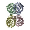
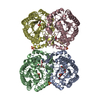
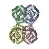
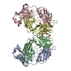
| Title | Crystal structure of phenylalanine-regulated 3-deoxy-D-arabino-heptulosonate-7-phosphate synthase (DAHP synthase) from E.coli complexed with Mn2+, PEP, and Phe | ||||||
 Components Components | 3-deoxy-D-arabino-heptulosonate-7-phosphate synthase | ||||||
 Keywords Keywords | LYASE / beta/alpha barrel / allosteric inhibition / feedback regulation / aromatic biosynthetic pathway | ||||||
| Function / homology |  Function and homology information Function and homology information3-deoxy-7-phosphoheptulonate synthase / 3-deoxy-7-phosphoheptulonate synthase activity / chorismate biosynthetic process / aromatic amino acid biosynthetic process / amino acid biosynthetic process / identical protein binding / cytoplasm / cytosol Similarity search - Function | ||||||
| Biological species |  | ||||||
| Method |  X-RAY DIFFRACTION / SYNCHROTRON / MAD / Resolution: 2.8 Å X-RAY DIFFRACTION / SYNCHROTRON / MAD / Resolution: 2.8 Å | ||||||
 Authors Authors | Shumilin, I.A. / Zhao, C. / Bauerle, R. / Kretsinger, R.H. | ||||||
 Citation Citation | Journal: J.Mol.Biol. / Year: 2002 Title: Allosteric inhibition of 3-deoxy-D-arabino-heptulosonate-7-phosphate synthase alters the coordination of both substrates. Authors: Shumilin, I.A. / Zhao, C. / Bauerle, R. / Kretsinger, R.H. | ||||||
| History |
|
- Structure visualization
| Structure viewer | Molecule: MolmilJmol/JSmol |
|---|
- Downloads & links
-Download
| PDBx/mmCIF format | 1kfl.cif.gz | 536.4 KB | Display | PDBx/mmCIF format |
|---|---|---|---|---|
| PDB format | pdb1kfl.ent.gz | 445.3 KB | Display | PDB format |
| PDBx/mmJSON format | 1kfl.json.gz | Tree view | PDBx/mmJSON format | |
| Others |  Other downloads Other downloads |
-Validation report
| Arichive directory | https://data.pdbj.org/pub/pdb/validation_reports/kf/1kflftp://data.pdbj.org/pub/pdb/validation_reports/kf/1kfl | HTTPS FTP |
|---|
-Related structure data
| Related structure data |  1qr7S S: Starting model for refinement |
|---|---|
| Similar structure data |

-Links
 PDBj
PDBj


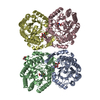
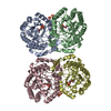
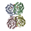
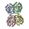
- Assembly
| Deposited unit | 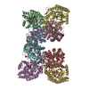
| ||||||||||
|---|---|---|---|---|---|---|---|---|---|---|---|
| 1 | 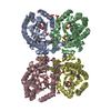
| ||||||||||
| 2 | 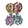
| ||||||||||
| Unit cell |
| ||||||||||
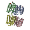
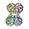
| Details | The biological assembly is a tetramer, two tetramers, ABCD and EFGH, reside in the asymmetric unit. |
-Components
| #1: Protein | Mass: 38434.660 Da / Num. of mol.: 8 Source method: isolated from a genetically manipulated source Source: (gene. exp.) References: UniProt: P00886, UniProt: P0AB91*PLUS, EC: 4.1.2.15 #2: Chemical | ChemComp-MN /   Mass: 54.938 Da / Num. of mol.: 8 / Source method: obtained synthetically / Formula: Mn Mass: 54.938 Da / Num. of mol.: 8 / Source method: obtained synthetically / Formula: Mn#3: Chemical | ChemComp-SO4 /   Mass: 96.063 Da / Num. of mol.: 16 / Source method: obtained synthetically / Formula: SO4 Mass: 96.063 Da / Num. of mol.: 16 / Source method: obtained synthetically / Formula: SO4#4: Chemical | ChemComp-PHE /   Type: L-peptide linking / Mass: 165.189 Da / Num. of mol.: 8 / Source method: obtained synthetically / Formula: C9H11NO2 Type: L-peptide linking / Mass: 165.189 Da / Num. of mol.: 8 / Source method: obtained synthetically / Formula: C9H11NO2#5: Chemical | ChemComp-PEP / 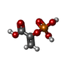  Mass: 168.042 Da / Num. of mol.: 8 / Source method: obtained synthetically / Formula: C3H5O6P Mass: 168.042 Da / Num. of mol.: 8 / Source method: obtained synthetically / Formula: C3H5O6PHas protein modification | Y | |
|---|
-Experimental details
-Experiment
| Experiment | Method: X-RAY DIFFRACTION / Number of used crystals: 1 |
|---|
- Sample preparation
| Crystal | Density Matthews: 2.84 Å3/Da / Density % sol: 56.75 % | ||||||||||||||||||||||||||||||||||||||||||||||||||||||||
|---|---|---|---|---|---|---|---|---|---|---|---|---|---|---|---|---|---|---|---|---|---|---|---|---|---|---|---|---|---|---|---|---|---|---|---|---|---|---|---|---|---|---|---|---|---|---|---|---|---|---|---|---|---|---|---|---|---|
| Crystal grow | Temperature: 295 K / Method: vapor diffusion, hanging drop / pH: 8.1 Details: PEG 4000, lithium sulfate, manganese sulfate, phosphoenolpyruvate, phenylalanine, bis-tris-propane, pH 8.1, VAPOR DIFFUSION, HANGING DROP at 295K | ||||||||||||||||||||||||||||||||||||||||||||||||||||||||
| Crystal | *PLUS Density % sol: 58 % | ||||||||||||||||||||||||||||||||||||||||||||||||||||||||
| Crystal grow | *PLUS | ||||||||||||||||||||||||||||||||||||||||||||||||||||||||
| Components of the solutions | *PLUS
|
-Data collection
| Diffraction | Mean temperature: 200 K |
|---|---|
| Diffraction source | Source: SYNCHROTRON / Site: NSLS  / Beamline: X4A / Wavelength: 0.97165 Å / Beamline: X4A / Wavelength: 0.97165 Å |
| Detector | Type: ADSC QUANTUM 4 / Detector: CCD / Date: Aug 15, 2000 / Details: mirrors |
| Radiation | Monochromator: Sagitally focused Si(111) / Protocol: MAD / Monochromatic (M) / Laue (L): M / Scattering type: x-ray |
| Radiation wavelength | Wavelength: 0.97165 Å / Relative weight: 1 |
| Reflection | Resolution: 2.8→20 Å / Num. all: 84564 / Num. obs: 84546 / % possible obs: 99.3 % / Observed criterion σ(F): 0 / Observed criterion σ(I): 0 / Redundancy: 6.8 % / Biso Wilson estimate: 38.6 Å2 / Rmerge(I) obs: 0.076 / Net I/σ(I): 24 |
| Reflection shell | Resolution: 2.8→2.9 Å / Redundancy: 6.5 % / Rmerge(I) obs: 0.313 / Mean I/σ(I) obs: 5.3 / % possible all: 98.7 |
| Reflection | *PLUS Highest resolution: 2.8 Å / Num. obs: 84564 / % possible obs: 99.4 % / Rmerge(I) obs: 0.076 |
| Reflection shell | *PLUS % possible obs: 98.7 % / Rmerge(I) obs: 0.313 |
- Processing
| Software |
| ||||||||||||||||||||||||||||||
|---|---|---|---|---|---|---|---|---|---|---|---|---|---|---|---|---|---|---|---|---|---|---|---|---|---|---|---|---|---|---|---|
| Refinement | Method to determine structure: MAD Starting model: PDB ENTRY 1QR7 Resolution: 2.8→20 Å / Isotropic thermal model: Anisotropic / Cross valid method: THROUGHOUT / σ(F): 0 / Stereochemistry target values: Engh & Huber
| ||||||||||||||||||||||||||||||
| Displacement parameters |
| ||||||||||||||||||||||||||||||
| Refinement step | Cycle: LAST / Resolution: 2.8→20 Å
| ||||||||||||||||||||||||||||||
| Refinement | *PLUS % reflection Rfree: 3 % / Rfactor all: 0.219 / Rfactor Rfree: 0.246 / Rfactor Rwork: 0.218 | ||||||||||||||||||||||||||||||
| Solvent computation | *PLUS | ||||||||||||||||||||||||||||||
| Displacement parameters | *PLUS | ||||||||||||||||||||||||||||||
| Refine LS restraints | *PLUS
|
