 Movie
Movie Controller

[English] 日本語
 Yorodumi
- PDB-5cks: DAHP (3-deoxy-D-arabinoheptulosonate-7-phosphate) Synthase in com... -
+ Open data

- Basic information
| Entry | Database: PDB / ID: 5cks | |||||||||
|---|---|---|---|---|---|---|---|---|---|---|
| Title | DAHP (3-deoxy-D-arabinoheptulosonate-7-phosphate) Synthase in complex with DAHP Oxime. | |||||||||
 Components Components | Phospho-2-dehydro-3-deoxyheptonate aldolase, Phe-sensitive | |||||||||
 Keywords Keywords | Transferase/transferase inhibitor / DAHP synthase / Inhibitor / Complex / Transition state mimic / Transferase-transferase inhibitor complex | |||||||||
| Function / homology |  Function and homology information Function and homology information3-deoxy-7-phosphoheptulonate synthase / 3-deoxy-7-phosphoheptulonate synthase activity / chorismate biosynthetic process / aromatic amino acid biosynthetic process / amino acid biosynthetic process / identical protein binding / cytosol / cytoplasm Similarity search - Function | |||||||||
| Biological species |  | |||||||||
| Method |  X-RAY DIFFRACTION / SYNCHROTRON / MOLECULAR REPLACEMENT / Resolution: 2.1181 Å X-RAY DIFFRACTION / SYNCHROTRON / MOLECULAR REPLACEMENT / Resolution: 2.1181 Å | |||||||||
 Authors Authors | Berti, P. / Junop, M. / Balachandran, N. | |||||||||
| Funding support |  Canada, 2items Canada, 2items
| |||||||||
 Citation Citation | Journal: Biochemistry / Year: 2016 Title: Potent Inhibition of 3-Deoxy-d-arabinoheptulosonate-7-phosphate (DAHP) Synthase by DAHP Oxime, a Phosphate Group Mimic. Authors: Balachandran, N. / Heimhalt, M. / Liuni, P. / To, F. / Wilson, D.J. / Junop, M.S. / Berti, P.J. | |||||||||
| History |
|
- Structure visualization
| Structure viewer | Molecule: MolmilJmol/JSmol |
|---|
- Downloads & links
-Download
| PDBx/mmCIF format | 5cks.cif.gz | 284.5 KB | Display | PDBx/mmCIF format |
|---|---|---|---|---|
| PDB format | pdb5cks.ent.gz | 228.9 KB | Display | PDB format |
| PDBx/mmJSON format | 5cks.json.gz | Tree view | PDBx/mmJSON format | |
| Others |  Other downloads Other downloads |
-Validation report
| Arichive directory | https://data.pdbj.org/pub/pdb/validation_reports/ck/5cksftp://data.pdbj.org/pub/pdb/validation_reports/ck/5cks | HTTPS FTP |
|---|
-Related structure data
| Related structure data | 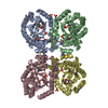 1kflS S: Starting model for refinement |
|---|---|
| Similar structure data |

-Links
 PDBj
PDBj

- Assembly
| Deposited unit | 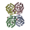
| ||||||||
|---|---|---|---|---|---|---|---|---|---|
| 1 |
| ||||||||
| Unit cell |
| ||||||||
| Components on special symmetry positions |
| ||||||||
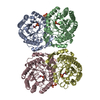
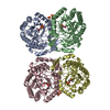
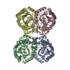
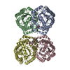
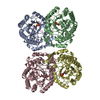
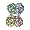
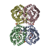
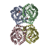
| Details | Tetramer confirmed by gel filtration |
-Components
| #1: Protein | Mass: 38116.555 Da / Num. of mol.: 4 Source method: isolated from a genetically manipulated source Details: first 5 residues are unstructured in the crystal structure. Also loops 101-102 and 313-317. Source: (gene. exp.) Strain: K12 / Gene: aroG, b0754, JW0737 / Plasmid: pET300 / Cell (production host): BL21(DE3) / Production host: References: UniProt: P0AB91, 3-deoxy-7-phosphoheptulonate synthase #2: Sugar |   Type: D-saccharide, beta linking / Mass: 180.156 Da / Num. of mol.: 3 Type: D-saccharide, beta linking / Mass: 180.156 Da / Num. of mol.: 3Source method: isolated from a genetically manipulated source Formula: C6H12O6 #3: Chemical | 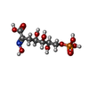  Mass: 303.161 Da / Num. of mol.: 2 / Source method: obtained synthetically / Formula: C7H14NO10P Mass: 303.161 Da / Num. of mol.: 2 / Source method: obtained synthetically / Formula: C7H14NO10P#4: Water | ChemComp-HOH / |  Mass: 18.015 Da / Num. of mol.: 833 / Source method: isolated from a natural source / Formula: H2O Mass: 18.015 Da / Num. of mol.: 833 / Source method: isolated from a natural source / Formula: H2O |
|---|
-Experimental details
-Experiment
| Experiment | Method: X-RAY DIFFRACTION |
|---|
- Sample preparation
| Crystal | Density Matthews: 2.5 Å3/Da / Density % sol: 50.79 % |
|---|---|
| Crystal grow | Temperature: 277 K / Method: vapor diffusion, hanging drop Details: Mother liquor (20% PEG 3350, 200 mM trilithium citrate tetrahydrate, 6% galactose) was mixed in equal volume with DHAPS at 10 mg/ml in buffer containing 20 mM Tris-HCl, 0.1 mM TCEP. PH range: 7 |
-Data collection
| Diffraction | Mean temperature: 100 K |
|---|---|
| Diffraction source | Source: SYNCHROTRON / Site: NSLS  / Beamline: X25 / Wavelength: 1.1 Å / Beamline: X25 / Wavelength: 1.1 Å |
| Detector | Type: DECTRIS PILATUS 6M / Detector: PIXEL / Date: Apr 28, 2010 |
| Radiation | Protocol: SINGLE WAVELENGTH / Monochromatic (M) / Laue (L): M / Scattering type: x-ray |
| Radiation wavelength | Wavelength: 1.1 Å / Relative weight: 1 |
| Reflection | Resolution: 2.1181→50 Å / Num. all: 85303 / Num. obs: 85303 / % possible obs: 99.9 % / Redundancy: 3.7 % / Rmerge(I) obs: 0.093 / Net I/σ(I): 12.8 |
| Reflection shell | Resolution: 2.1181→2.16 Å / Redundancy: 3.6 % / Rmerge(I) obs: 0.406 / Mean I/σ(I) obs: 3.3 / % possible all: 100 |
- Processing
| Software |
| |||||||||||||||||||||||||||||||||||||||||||||||||||||||||||||||||||||||||||||||||||||||||||||||||||||||||
|---|---|---|---|---|---|---|---|---|---|---|---|---|---|---|---|---|---|---|---|---|---|---|---|---|---|---|---|---|---|---|---|---|---|---|---|---|---|---|---|---|---|---|---|---|---|---|---|---|---|---|---|---|---|---|---|---|---|---|---|---|---|---|---|---|---|---|---|---|---|---|---|---|---|---|---|---|---|---|---|---|---|---|---|---|---|---|---|---|---|---|---|---|---|---|---|---|---|---|---|---|---|---|---|---|---|---|
| Refinement | Method to determine structure: MOLECULAR REPLACEMENT Starting model: 1KFL Resolution: 2.1181→42.424 Å / SU ML: 0.25 / Cross valid method: FREE R-VALUE / σ(F): 1.34 / Phase error: 24 / Stereochemistry target values: ML
| |||||||||||||||||||||||||||||||||||||||||||||||||||||||||||||||||||||||||||||||||||||||||||||||||||||||||
| Solvent computation | Shrinkage radii: 0.86 Å / VDW probe radii: 1.1 Å / Solvent model: FLAT BULK SOLVENT MODEL / Bsol: 36.415 Å2 / ksol: 0.339 e/Å3 | |||||||||||||||||||||||||||||||||||||||||||||||||||||||||||||||||||||||||||||||||||||||||||||||||||||||||
| Displacement parameters | Biso mean: 30.6 Å2
| |||||||||||||||||||||||||||||||||||||||||||||||||||||||||||||||||||||||||||||||||||||||||||||||||||||||||
| Refinement step | Cycle: LAST / Resolution: 2.1181→42.424 Å
| |||||||||||||||||||||||||||||||||||||||||||||||||||||||||||||||||||||||||||||||||||||||||||||||||||||||||
| Refine LS restraints |
| |||||||||||||||||||||||||||||||||||||||||||||||||||||||||||||||||||||||||||||||||||||||||||||||||||||||||
| LS refinement shell |
|
