 Movie
Movie Controller

[English] 日本語
 Yorodumi
- PDB-1n8f: Crystal structure of E24Q mutant of phenylalanine-regulated 3-deo... -
+ Open data

- Basic information
| Entry | Database: PDB / ID: 1n8f | ||||||
|---|---|---|---|---|---|---|---|
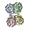
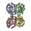
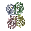
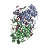
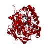
| Title | Crystal structure of E24Q mutant of phenylalanine-regulated 3-deoxy-D-arabino-heptulosonate-7-phosphate synthase (DAHP synthase) from Escherichia Coli in complex with Mn2+ and PEP | ||||||
 Components Components | DAHP Synthetase | ||||||
 Keywords Keywords | METAL BINDING PROTEIN / (Beta/Alpha)8 Barrel | ||||||
| Function / homology |  Function and homology information Function and homology information3-deoxy-7-phosphoheptulonate synthase / 3-deoxy-7-phosphoheptulonate synthase activity / chorismate biosynthetic process / aromatic amino acid biosynthetic process / amino acid biosynthetic process / identical protein binding / cytosol / cytoplasm Similarity search - Function | ||||||
| Biological species |  | ||||||
| Method |  X-RAY DIFFRACTION / SYNCHROTRON / MOLECULAR REPLACEMENT / Resolution: 1.75 Å X-RAY DIFFRACTION / SYNCHROTRON / MOLECULAR REPLACEMENT / Resolution: 1.75 Å | ||||||
 Authors Authors | Shumilin, I.A. / Bauerle, R. / Kretsinger, R.H. | ||||||
 Citation Citation | Journal: Biochemistry / Year: 2003 Title: The High-Resolution Structure of 3-Deoxy-D-arabino-heptulosonate-7-phosphate Synthase Reveals a Twist in the Plane of Bound Phosphoenolpyruvate Authors: Shumilin, I.A. / Bauerle, R. / Kretsinger, R.H. | ||||||
| History |
|
- Structure visualization
| Structure viewer | Molecule: MolmilJmol/JSmol |
|---|
- Downloads & links
-Download
| PDBx/mmCIF format | 1n8f.cif.gz | 299.7 KB | Display | PDBx/mmCIF format |
|---|---|---|---|---|
| PDB format | pdb1n8f.ent.gz | 239.6 KB | Display | PDB format |
| PDBx/mmJSON format | 1n8f.json.gz | Tree view | PDBx/mmJSON format | |
| Others |  Other downloads Other downloads |
-Validation report
| Arichive directory | https://data.pdbj.org/pub/pdb/validation_reports/n8/1n8fftp://data.pdbj.org/pub/pdb/validation_reports/n8/1n8f | HTTPS FTP |
|---|
-Related structure data
| Related structure data | |
|---|---|
| Similar structure data |


-Links
 PDBj
PDBj
- Assembly
| Deposited unit | 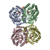
| ||||||||
|---|---|---|---|---|---|---|---|---|---|
| 1 | 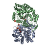
| ||||||||
| 2 | 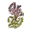
| ||||||||
| Unit cell |
| ||||||||
| Details | The biological assembly of the wild type DAHP synthase is a tetramer similar to the one located in the asymmetric unit of E24Q mutant. |
-Components
| #1: Protein | Mass: 38058.520 Da / Num. of mol.: 4 / Mutation: E24Q Source method: isolated from a genetically manipulated source Source: (gene. exp.) #2: Chemical | ChemComp-MN /   Mass: 54.938 Da / Num. of mol.: 4 / Source method: obtained synthetically / Formula: Mn Mass: 54.938 Da / Num. of mol.: 4 / Source method: obtained synthetically / Formula: Mn#3: Chemical | ChemComp-SO4 /   Mass: 96.063 Da / Num. of mol.: 8 / Source method: obtained synthetically / Formula: SO4 Mass: 96.063 Da / Num. of mol.: 8 / Source method: obtained synthetically / Formula: SO4#4: Chemical | ChemComp-PEP /   Mass: 168.042 Da / Num. of mol.: 4 / Source method: obtained synthetically / Formula: C3H5O6P Mass: 168.042 Da / Num. of mol.: 4 / Source method: obtained synthetically / Formula: C3H5O6P#5: Water | ChemComp-HOH / |  Mass: 18.015 Da / Num. of mol.: 1395 / Source method: isolated from a natural source / Formula: H2O Mass: 18.015 Da / Num. of mol.: 1395 / Source method: isolated from a natural source / Formula: H2O |
|---|
-Experimental details
-Experiment
| Experiment | Method: X-RAY DIFFRACTION / Number of used crystals: 1 |
|---|
- Sample preparation
| Crystal | Density Matthews: 2.28 Å3/Da / Density % sol: 45.57 % | |||||||||||||||||||||||||||||||||||||||||||||||||||||||||||||||||||||||||||||
|---|---|---|---|---|---|---|---|---|---|---|---|---|---|---|---|---|---|---|---|---|---|---|---|---|---|---|---|---|---|---|---|---|---|---|---|---|---|---|---|---|---|---|---|---|---|---|---|---|---|---|---|---|---|---|---|---|---|---|---|---|---|---|---|---|---|---|---|---|---|---|---|---|---|---|---|---|---|---|
| Crystal grow | Temperature: 294 K / Method: vapor diffusion, hanging drop / pH: 8.1 Details: PEG4000, manganese sulfate, phosphoenolpyruvate, lithium sulfate, bis-tris propane, pH 8.1, VAPOR DIFFUSION, HANGING DROP, temperature 294K | |||||||||||||||||||||||||||||||||||||||||||||||||||||||||||||||||||||||||||||
| Crystal grow | *PLUS | |||||||||||||||||||||||||||||||||||||||||||||||||||||||||||||||||||||||||||||
| Components of the solutions | *PLUS
|
-Data collection
| Diffraction | Mean temperature: 200 K |
|---|---|
| Diffraction source | Source: SYNCHROTRON / Site: NSLS  / Beamline: X4A / Wavelength: 0.9787 Å / Beamline: X4A / Wavelength: 0.9787 Å |
| Detector | Type: ADSC QUANTUM 4 / Detector: CCD / Date: Aug 19, 2001 / Details: mirrors |
| Radiation | Monochromator: SAGITALLY FOCUSED Si(111) / Protocol: SINGLE WAVELENGTH / Monochromatic (M) / Laue (L): M / Scattering type: x-ray |
| Radiation wavelength | Wavelength: 0.9787 Å / Relative weight: 1 |
| Reflection | Resolution: 1.75→20 Å / Num. all: 152257 / Num. obs: 151346 / % possible obs: 99.4 % / Observed criterion σ(I): -3 / Redundancy: 4.2 % / Biso Wilson estimate: 21.5 Å2 / Rmerge(I) obs: 0.059 / Net I/σ(I): 15.5 |
| Reflection shell | Resolution: 1.75→1.81 Å / Redundancy: 3.8 % / Rmerge(I) obs: 0.348 / Mean I/σ(I) obs: 2.5 / % possible all: 98.6 |
| Reflection shell | *PLUS % possible obs: 98.6 % |
- Processing
| Software |
| |||||||||||||||||||||||||||
|---|---|---|---|---|---|---|---|---|---|---|---|---|---|---|---|---|---|---|---|---|---|---|---|---|---|---|---|---|
| Refinement | Method to determine structure: MOLECULAR REPLACEMENT / Resolution: 1.75→20 Å / Cross valid method: THROUGHOUT / σ(F): 0 / Stereochemistry target values: Engh & Huber
| |||||||||||||||||||||||||||
| Displacement parameters | Biso mean: 31.3 Å2
| |||||||||||||||||||||||||||
| Refine analyze |
| |||||||||||||||||||||||||||
| Refinement step | Cycle: LAST / Resolution: 1.75→20 Å
| |||||||||||||||||||||||||||
| Refine LS restraints |
| |||||||||||||||||||||||||||
| Refinement | *PLUS % reflection Rfree: 2 % | |||||||||||||||||||||||||||
| Solvent computation | *PLUS | |||||||||||||||||||||||||||
| Displacement parameters | *PLUS | |||||||||||||||||||||||||||
| Refine LS restraints | *PLUS
|
