Movie
Movie Controller

[English] 日本語
 Yorodumi
- EMDB-3443: Electron cryo-microscopy of the yeast RNA polymerase I - Rrn3 com... -
+ Open data

- Basic information
| Entry | Database: EMDB / ID: EMD-3443 | |||||||||
|---|---|---|---|---|---|---|---|---|---|---|
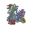
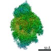
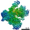
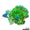
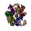
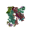
| Title | Electron cryo-microscopy of the yeast RNA polymerase I - Rrn3 complex at 7.5A resolution | |||||||||
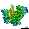
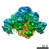
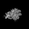
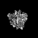
 Map data Map data | reconstruction of the RNA pol I - Rrn3 complex | |||||||||
 Sample Sample |
| |||||||||
 Keywords Keywords | RNA polymerase I initiation factor Rrn3 transcription initiation | |||||||||
| Biological species |  | |||||||||
| Method | single particle reconstruction / cryo EM / negative staining / Resolution: 7.5 Å | |||||||||
 Authors Authors | Pilsl M / Crucifix C / Papai G / Krupp F / Steinbauer R / Griesenbeck J / Milkereit P / Tschochner H / Schultz P | |||||||||
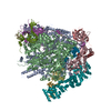
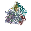
 Citation Citation | Journal: Nat Commun / Year: 2016 Title: Structure of the initiation-competent RNA polymerase I and its implication for transcription. Authors: Michael Pilsl / Corinne Crucifix / Gabor Papai / Ferdinand Krupp / Robert Steinbauer / Joachim Griesenbeck / Philipp Milkereit / Herbert Tschochner / Patrick Schultz /   Abstract: Eukaryotic RNA polymerase I (Pol I) is specialized in rRNA gene transcription synthesizing up to 60% of cellular RNA. High level rRNA production relies on efficient binding of initiation factors to ...Eukaryotic RNA polymerase I (Pol I) is specialized in rRNA gene transcription synthesizing up to 60% of cellular RNA. High level rRNA production relies on efficient binding of initiation factors to the rRNA gene promoter and recruitment of Pol I complexes containing initiation factor Rrn3. Here, we determine the cryo-EM structure of the Pol I-Rrn3 complex at 7.5 Å resolution, and compare it with Rrn3-free monomeric and dimeric Pol I. We observe that Rrn3 contacts the Pol I A43/A14 stalk and subunits A190 and AC40, that association re-organizes the Rrn3 interaction interface, thereby preventing Pol I dimerization; and Rrn3-bound and monomeric Pol I differ from the dimeric enzyme in cleft opening, and localization of the A12.2 C-terminus in the active centre. Our findings thus support a dual role for Rrn3 in transcription initiation to stabilize a monomeric initiation competent Pol I and to drive pre-initiation complex formation. | |||||||||
| History |
|
- Structure visualization
| Movie |
 Movie viewer Movie viewer |
|---|---|
| Structure viewer | EM map: SurfViewMolmilJmol/JSmol |
| Supplemental images |

- Downloads & links
-EMDB archive
| Map data | emd_3443.map.gz | 7.4 MB | EMDB map data format | |
|---|---|---|---|---|
| Header (meta data) | emd-3443-v30.xmlemd-3443.xml | 10.6 KB 10.6 KB | Display Display | EMDB header |
| Images |  emd_3443.png emd_3443.png | 352.9 KB | ||
| Archive directory |  http://ftp.pdbj.org/pub/emdb/structures/EMD-3443ftp://ftp.pdbj.org/pub/emdb/structures/EMD-3443 http://ftp.pdbj.org/pub/emdb/structures/EMD-3443ftp://ftp.pdbj.org/pub/emdb/structures/EMD-3443 | HTTPS FTP |
-Related structure data
| Similar structure data |
|---|
-Links
| EMDB pages | EMDB (EBI/PDBe) / EMDataResource |
|---|
-Map
| File | Download / File: emd_3443.map.gz / Format: CCP4 / Size: 7.8 MB / Type: IMAGE STORED AS FLOATING POINT NUMBER (4 BYTES) | ||||||||||||||||||||||||||||||||||||||||||||||||||||||||||||||||||||
|---|---|---|---|---|---|---|---|---|---|---|---|---|---|---|---|---|---|---|---|---|---|---|---|---|---|---|---|---|---|---|---|---|---|---|---|---|---|---|---|---|---|---|---|---|---|---|---|---|---|---|---|---|---|---|---|---|---|---|---|---|---|---|---|---|---|---|---|---|---|
| Annotation | reconstruction of the RNA pol I - Rrn3 complex | ||||||||||||||||||||||||||||||||||||||||||||||||||||||||||||||||||||
| Projections & slices | Image control
Images are generated by Spider. | ||||||||||||||||||||||||||||||||||||||||||||||||||||||||||||||||||||
| Voxel size | X=Y=Z: 2.16 Å | ||||||||||||||||||||||||||||||||||||||||||||||||||||||||||||||||||||
| Density |
| ||||||||||||||||||||||||||||||||||||||||||||||||||||||||||||||||||||
| Symmetry | Space group: 1 | ||||||||||||||||||||||||||||||||||||||||||||||||||||||||||||||||||||
| Details | EMDB XML:
CCP4 map header:
| ||||||||||||||||||||||||||||||||||||||||||||||||||||||||||||||||||||
 Z (Sec.)
 Y (Row.)
 X (Col.)


-Supplemental data
- Sample components
-Entire : complex formed between YEAST RNA polymerase I and the initiation ...
| Entire | Name: complex formed between YEAST RNA polymerase I and the initiation factor Rrn3 |
|---|---|
| Components |
|
-Supramolecule #1000: complex formed between YEAST RNA polymerase I and the initiation ...
| Supramolecule | Name: complex formed between YEAST RNA polymerase I and the initiation factor Rrn3 type: sample / ID: 1000 / Oligomeric state: 1 / Number unique components: 2 |
|---|---|
| Molecular weight | Theoretical: 663 MDa |
-Macromolecule #1: DNA dependent RNA polymerase I
| Macromolecule | Name: DNA dependent RNA polymerase I / type: protein_or_peptide / ID: 1 / Number of copies: 1 / Oligomeric state: monomer / Recombinant expression: No |
|---|---|
| Source (natural) | Organism: |
| Molecular weight | Theoretical: 590 KDa |
-Macromolecule #2: Rrn3
| Macromolecule | Name: Rrn3 / type: protein_or_peptide / ID: 2 / Number of copies: 1 / Oligomeric state: monomer / Recombinant expression: No |
|---|---|
| Source (natural) | Organism: |
| Molecular weight | Theoretical: 72.346 KDa |
-Experimental details
-Structure determination
| Method | negative staining, cryo EM |
|---|---|
 Processing Processing | single particle reconstruction |
| Aggregation state | particle |
-Sample preparation
| Concentration | 0.01 mg/mL |
|---|---|
| Buffer | pH: 7.8 / Details: 20 mM Hepes, 100mM ammonium acetate and 2mM MgCl2 |
| Staining | Type: NEGATIVE / Details: unstained |
| Grid | Details: sample was adsorbed on a floated carbon foil which was deposited onto a quantifoil grid |
| Vitrification | Cryogen name: ETHANE / Chamber humidity: 95 % / Instrument: FEI VITROBOT MARK IV / Method: blotting time (4 sec.) blotting force 5 |
- Electron microscopy
| Microscope | FEI TITAN KRIOS |
|---|---|
| Temperature | Min: 80 K / Max: 100 K / Average: 90 K |
| Alignment procedure | Legacy - Astigmatism: Cs corrector |
| Date | Jun 10, 2015 |
| Image recording | Category: CCD / Film or detector model: FEI FALCON II (4k x 4k) / Average electron dose: 22 e/Å2 |
| Electron beam | Acceleration voltage: 300 kV / Electron source:  FIELD EMISSION GUN FIELD EMISSION GUN |
| Electron optics | Calibrated magnification: 129630 / Illumination mode: SPOT SCAN / Imaging mode: BRIGHT FIELD / Cs: 0.01 mm / Nominal defocus max: 3.5 µm / Nominal defocus min: 1.5 µm / Nominal magnification: 59000 |
| Sample stage | Specimen holder model: FEI TITAN KRIOS AUTOGRID HOLDER |
| Experimental equipment |  Model: Titan Krios / Image courtesy: FEI Company |
-Image processing
| CTF correction | Details: each particle |
|---|---|
| Final reconstruction | Applied symmetry - Point group: C1 (asymmetric) / Algorithm: OTHER / Resolution.type: BY AUTHOR / Resolution: 7.5 Å / Resolution method: OTHER / Software - Name: relion / Details: final map was calculated from 2 averaged datasets / Number images used: 32438 |
-Atomic model buiding 1
| Initial model | PDB ID: |
|---|---|
| Software | Name: gEMfitter, Chimera |
| Refinement | Space: REAL / Protocol: RIGID BODY FIT |