Movie
Movie Controller


+ Open data

- Basic information
| Entry | Database: PDB / ID: 5g5l | ||||||
|---|---|---|---|---|---|---|---|
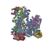
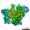
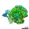
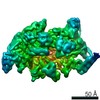
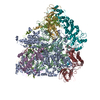
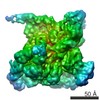
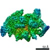
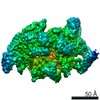
| Title | RNA polymerase I-Rrn3 complex at 4.8 A resolution | ||||||
 Components Components |
| ||||||
 Keywords Keywords | RNA POLYMERASE / TRANSCIPTION | ||||||
| Function / homology |  Function and homology information Function and homology informationRNA polymerase I general transcription initiation factor activity / RNA polymerase I core binding / RNA polymerase I general transcription initiation factor binding / nuclear DNA-directed RNA polymerase complex / rDNA binding / RNA polymerase I preinitiation complex assembly / RNA Polymerase I Transcription Initiation / regulation of cell size / Processing of Capped Intron-Containing Pre-mRNA / RNA Polymerase III Transcription Initiation From Type 2 Promoter ...RNA polymerase I general transcription initiation factor activity / RNA polymerase I core binding / RNA polymerase I general transcription initiation factor binding / nuclear DNA-directed RNA polymerase complex / rDNA binding / RNA polymerase I preinitiation complex assembly / RNA Polymerase I Transcription Initiation / regulation of cell size / Processing of Capped Intron-Containing Pre-mRNA / RNA Polymerase III Transcription Initiation From Type 2 Promoter / RNA Pol II CTD phosphorylation and interaction with CE / Formation of the Early Elongation Complex / mRNA Capping / RNA polymerase II transcribes snRNA genes / TP53 Regulates Transcription of DNA Repair Genes / RNA Polymerase II Promoter Escape / RNA Polymerase II Transcription Pre-Initiation And Promoter Opening / RNA Polymerase II Transcription Initiation / RNA Polymerase II Transcription Initiation And Promoter Clearance / RNA Polymerase II Pre-transcription Events / Formation of TC-NER Pre-Incision Complex / RNA-templated transcription / RNA Polymerase I Promoter Escape / termination of RNA polymerase III transcription / termination of RNA polymerase I transcription / transcription initiation at RNA polymerase III promoter / Gap-filling DNA repair synthesis and ligation in TC-NER / nucleolar large rRNA transcription by RNA polymerase I / transcription initiation at RNA polymerase I promoter / Estrogen-dependent gene expression / transcription by RNA polymerase III / Dual incision in TC-NER / RNA polymerase I complex / RNA polymerase III complex / transcription elongation by RNA polymerase I / RNA polymerase II, core complex / tRNA transcription by RNA polymerase III / transcription by RNA polymerase I / promoter-specific chromatin binding / transcription initiation at RNA polymerase II promoter / transcription elongation by RNA polymerase II / ribonucleoside binding / DNA-directed RNA polymerase / DNA-directed RNA polymerase activity / peroxisome / ribosome biogenesis / transcription by RNA polymerase II / nucleic acid binding / RNA polymerase II-specific DNA-binding transcription factor binding / protein dimerization activity / nucleolus / negative regulation of transcription by RNA polymerase II / DNA binding / zinc ion binding / nucleoplasm / metal ion binding / nucleus / cytoplasm Similarity search - Function | ||||||
| Biological species |  | ||||||
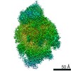
| Method | ELECTRON MICROSCOPY / single particle reconstruction / cryo EM / Resolution: 4.8 Å | ||||||
 Authors Authors | Engel, C. / Plitzko, J. / Cramer, P. | ||||||
 Citation Citation | Journal: Nat Commun / Year: 2016 Title: RNA polymerase I-Rrn3 complex at 4.8 Å resolution. Authors: Christoph Engel / Jürgen Plitzko / Patrick Cramer /  Abstract: Transcription of ribosomal DNA by RNA polymerase I (Pol I) requires the initiation factor Rrn3. Here we report the cryo-EM structure of the Pol I-Rrn3 complex at 4.8 Å resolution. The structure ...Transcription of ribosomal DNA by RNA polymerase I (Pol I) requires the initiation factor Rrn3. Here we report the cryo-EM structure of the Pol I-Rrn3 complex at 4.8 Å resolution. The structure reveals how Rrn3 binding converts an inactive Pol I dimer into an initiation-competent monomeric complex and provides insights into the mechanisms of Pol I-specific initiation and regulation. | ||||||
| History |
| ||||||
| Remark 700 | SHEET DETERMINATION METHOD: DSSP THE SHEETS PRESENTED AS "AE" IN EACH CHAIN ON SHEET RECORDS BELOW ... SHEET DETERMINATION METHOD: DSSP THE SHEETS PRESENTED AS "AE" IN EACH CHAIN ON SHEET RECORDS BELOW IS ACTUALLY AN 6-STRANDED BARREL THIS IS REPRESENTED BY A 7-STRANDED SHEET IN WHICH THE FIRST AND LAST STRANDS ARE IDENTICAL. THE SHEETS PRESENTED AS "AH" IN EACH CHAIN ON SHEET RECORDS BELOW IS ACTUALLY AN 9-STRANDED BARREL THIS IS REPRESENTED BY A 10-STRANDED SHEET IN WHICH THE FIRST AND LAST STRANDS ARE IDENTICAL. THE SHEETS PRESENTED AS "BN" IN EACH CHAIN ON SHEET RECORDS BELOW IS ACTUALLY AN -4-STRANDED BARREL THIS IS REPRESENTED BY A -3-STRANDED SHEET IN WHICH THE FIRST AND LAST STRANDS ARE IDENTICAL. THE SHEETS PRESENTED AS "GC" IN EACH CHAIN ON SHEET RECORDS BELOW IS ACTUALLY AN -1-STRANDED BARREL THIS IS REPRESENTED BY A 0-STRANDED SHEET IN WHICH THE FIRST AND LAST STRANDS ARE IDENTICAL. THE SHEETS PRESENTED AS "MB" IN EACH CHAIN ON SHEET RECORDS BELOW IS ACTUALLY AN -3-STRANDED BARREL THIS IS REPRESENTED BY A -2-STRANDED SHEET IN WHICH THE FIRST AND LAST STRANDS ARE IDENTICAL. |
- Structure visualization
| Movie |
Movie viewer |
|---|---|
| Structure viewer | Molecule: MolmilJmol/JSmol |
- Downloads & links
-Download
| PDBx/mmCIF format | 5g5l.cif.gz | 839.9 KB | Display | PDBx/mmCIF format |
|---|---|---|---|---|
| PDB format | pdb5g5l.ent.gz | 642.8 KB | Display | PDB format |
| PDBx/mmJSON format | 5g5l.json.gz | Tree view | PDBx/mmJSON format | |
| Others |  Other downloads Other downloads |
-Validation report
| Arichive directory | https://data.pdbj.org/pub/pdb/validation_reports/g5/5g5lftp://data.pdbj.org/pub/pdb/validation_reports/g5/5g5l | HTTPS FTP |
|---|
-Related structure data
| Related structure data | 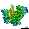 3439MC M: map data used to model this data C: citing same article ( |
|---|---|
| Similar structure data |
-Links
 PDBj
PDBj


- Assembly
| Deposited unit | 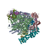
|
|---|---|
| 1 |
|
-Components
-DNA-DIRECTED RNA POLYMERASE I SUBUNIT ... , 7 types, 7 molecules ABDGIMN
| #1: Protein | Mass: 186676.969 Da / Num. of mol.: 1 / Source method: isolated from a natural source / Details: C-TERMINAL FLAG-10XHIS TAG / Source: (natural) |
|---|---|
| #2: Protein | Mass: 135910.328 Da / Num. of mol.: 1 / Source method: isolated from a natural source / Source: (natural) |
| #4: Protein | Mass: 14599.128 Da / Num. of mol.: 1 / Source method: isolated from a natural source / Source: (natural) |
| #7: Protein | Mass: 36264.852 Da / Num. of mol.: 1 / Source method: isolated from a natural source / Source: (natural) |
| #9: Protein | Mass: 13676.566 Da / Num. of mol.: 1 / Source method: isolated from a natural source / Source: (natural) |
| #13: Protein | Mass: 46721.707 Da / Num. of mol.: 1 / Source method: isolated from a natural source / Source: (natural) |
| #14: Protein | Mass: 26933.518 Da / Num. of mol.: 1 / Source method: isolated from a natural source / Source: (natural) |
-DNA-DIRECTED RNA POLYMERASES I AND III SUBUNIT ... , 2 types, 2 molecules CK
| #3: Protein | Mass: 37732.613 Da / Num. of mol.: 1 / Source method: isolated from a natural source / Source: (natural) |
|---|---|
| #11: Protein | Mass: 16167.860 Da / Num. of mol.: 1 / Source method: isolated from a natural source / Source: (natural) |
-DNA-DIRECTED RNA POLYMERASES I, II, AND III SUBUNIT RPABC ... , 5 types, 5 molecules EFHJL
| #5: Protein | Mass: 25117.094 Da / Num. of mol.: 1 / Source method: isolated from a natural source / Source: (natural) |
|---|---|
| #6: Protein | Mass: 17931.834 Da / Num. of mol.: 1 / Source method: isolated from a natural source / Source: (natural) |
| #8: Protein | Mass: 16525.363 Da / Num. of mol.: 1 / Source method: isolated from a natural source / Source: (natural) |
| #10: Protein | Mass: 8290.732 Da / Num. of mol.: 1 / Source method: isolated from a natural source / Source: (natural) |
| #12: Protein | Mass: 7729.969 Da / Num. of mol.: 1 / Source method: isolated from a natural source / Source: (natural) |
-Protein / Non-polymers , 2 types, 8 molecules O
| #15: Protein | Mass: 72458.258 Da / Num. of mol.: 1 Source method: isolated from a genetically manipulated source Source: (gene. exp.) Strain: CB010 / Plasmid: PET28 / Production host:  |
|---|---|
| #16: Chemical | ChemComp-ZN /  Mass: 65.409 Da / Num. of mol.: 7 / Source method: obtained synthetically / Formula: Zn Mass: 65.409 Da / Num. of mol.: 7 / Source method: obtained synthetically / Formula: Zn |
-Details
| Has protein modification | Y |
|---|---|
| Sequence details | C-TERMINAL FLAG-10XHIS TAG |
-Experimental details
-Experiment
| Experiment | Method: ELECTRON MICROSCOPY |
|---|---|
| EM experiment | Aggregation state: PARTICLE / 3D reconstruction method: single particle reconstruction |
- Sample preparation
| Component | Name: S. CEREVISIAE RNA POLYMERASE I BOUND TO THE INITIATION FACTOR RRN3 Type: COMPLEX |
|---|---|
| Buffer solution | Name: 150MM NACL, 5MM HEPES, 1MM MGCL2, 0.01 MM ZNCL2, 5 MM DTT pH: 7.8 Details: 150MM NACL, 5MM HEPES, 1MM MGCL2, 0.01 MM ZNCL2, 5 MM DTT |
| Specimen | Conc.: 0.1 mg/ml / Embedding applied: NO / Shadowing applied: NO / Staining applied: NO / Vitrification applied: YES |
| Specimen support | Details: HOLEY CARBON |
| Vitrification | Instrument: FEI VITROBOT MARK IV / Cryogen name: ETHANE Details: VITROBOT MARK IV, METHOD- 4.5 MICROLITERS OF SAMPLE WAS APPLIED TO GLOW- DISCHARGED QUANTIFOIL R 2- 1 HOLEY CARBON GRIDS, WHICH WERE THEN BLOTTED FOR 8.5S AND PLUNGE- FROZEN IN LIQUID ETHANE. |
- Electron microscopy imaging
| Experimental equipment |  Model: Titan Krios / Image courtesy: FEI Company |
|---|---|
| Microscopy | Model: FEI TITAN KRIOS / Date: Mar 27, 2015 |
| Electron gun | Electron source:  FIELD EMISSION GUN / Accelerating voltage: 300 kV / Illumination mode: FLOOD BEAM FIELD EMISSION GUN / Accelerating voltage: 300 kV / Illumination mode: FLOOD BEAM |
| Electron lens | Mode: BRIGHT FIELD / Nominal magnification: 37000 X / Calibrated magnification: 37037 X / Nominal defocus max: 3600 nm / Nominal defocus min: 800 nm / Cs: 2.7 mm |
| Image recording | Electron dose: 40 e/Å2 / Film or detector model: GATAN K2 SUMMIT (4k x 4k) |
| Image scans | Num. digital images: 1174 |
- Processing
| EM software |
| ||||||||||||
|---|---|---|---|---|---|---|---|---|---|---|---|---|---|
| CTF correction | Details: RELION | ||||||||||||
| Symmetry | Point symmetry: C1 (asymmetric) | ||||||||||||
| 3D reconstruction | Method: RELION / Resolution: 4.8 Å / Num. of particles: 63445 / Nominal pixel size: 1.35 Å / Actual pixel size: 1.35 Å Details: SUBMISSION BASED ON EXPERIMENTAL DATA FROM EMDB EMD-3439. (DEPOSITION ID: 14566). Symmetry type: POINT | ||||||||||||
| Atomic model building | B value: 150 / Protocol: RIGID BODY FIT / Space: REAL / Details: METHOD--RIGID BODY REFINEMENT PROTOCOL--X-RAY | ||||||||||||
| Atomic model building | PDB-ID: 4C2M Accession code: 4C2M / Source name: PDB / Type: experimental model | ||||||||||||
| Refinement | Highest resolution: 4.8 Å | ||||||||||||
| Refinement step | Cycle: LAST / Highest resolution: 4.8 Å
|