 Movie
Movie Controller

[English] 日本語
 Yorodumi
- EMDB-0379: Structure of bacteriophage T7 lagging-strand DNA polymerase (D5A/... -
+ Open data

- Basic information
| Entry | Database: EMDB / ID: EMD-0379 | |||||||||
|---|---|---|---|---|---|---|---|---|---|---|
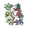
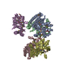
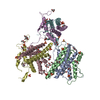
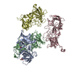
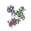
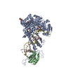
| Title | Structure of bacteriophage T7 lagging-strand DNA polymerase (D5A/E7A) interacting with primase domains of two gp4 subunits bound to an RNA/DNA hybrid and dTTP (from LagS1) | |||||||||
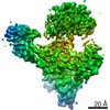
 Map data Map data | Structure of gp5 DNA polymerase and two gp4 primase subunits complexed with trx, RNA/DNA hybrid, and incoming dTTP (from gp4-gp5 lagging-strand complex LagS1) | |||||||||
 Sample Sample |
| |||||||||
 Keywords Keywords | DNA polymerase / primase / helicase / DNA replication / replisome / HYDROLASE / TRANSFERASE-DNA complex | |||||||||
| Function / homology |  Function and homology information Function and homology informationDNA synthesis involved in DNA replication / DNA exonuclease activity / DNA replication, synthesis of primer / viral DNA genome replication / Hydrolases; Acting on ester bonds; Exodeoxyribonucleases producing 5'-phosphomonoesters / 3'-5' exonuclease activity / DNA helicase activity / DNA-templated DNA replication / Transferases; Transferring phosphorus-containing groups; Nucleotidyltransferases / DNA-directed RNA polymerase activity ...DNA synthesis involved in DNA replication / DNA exonuclease activity / DNA replication, synthesis of primer / viral DNA genome replication / Hydrolases; Acting on ester bonds; Exodeoxyribonucleases producing 5'-phosphomonoesters / 3'-5' exonuclease activity / DNA helicase activity / DNA-templated DNA replication / Transferases; Transferring phosphorus-containing groups; Nucleotidyltransferases / DNA-directed RNA polymerase activity / double-strand break repair / single-stranded DNA binding / 5'-3' DNA helicase activity / DNA helicase / DNA-directed DNA polymerase / DNA-directed DNA polymerase activity / nucleotide binding / ATP hydrolysis activity / DNA binding / zinc ion binding / ATP binding / metal ion binding / identical protein binding Similarity search - Function | |||||||||
| Biological species |   Enterobacteria phage T7 (virus) Enterobacteria phage T7 (virus) | |||||||||
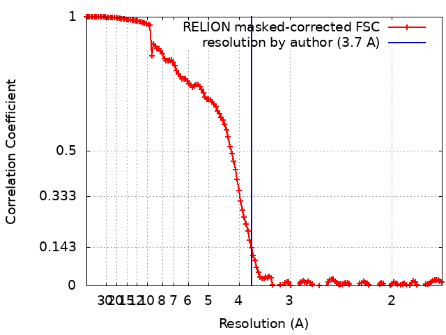
| Method | single particle reconstruction / cryo EM / Resolution: 3.7 Å | |||||||||
 Authors Authors | Gao Y / Fox T | |||||||||
| Funding support |  United States, 1 items United States, 1 items
| |||||||||
 Citation Citation | Journal: Science / Year: 2019 Title: Structures and operating principles of the replisome. Authors: Yang Gao / Yanxiang Cui / Tara Fox / Shiqiang Lin / Huaibin Wang / Natalia de Val / Z Hong Zhou / Wei Yang / Abstract: Visualization in atomic detail of the replisome that performs concerted leading- and lagging-DNA strand synthesis at a replication fork has not been reported. Using bacteriophage T7 as a model ...Visualization in atomic detail of the replisome that performs concerted leading- and lagging-DNA strand synthesis at a replication fork has not been reported. Using bacteriophage T7 as a model system, we determined cryo-electron microscopy structures up to 3.2-angstroms resolution of helicase translocating along DNA and of helicase-polymerase-primase complexes engaging in synthesis of both DNA strands. Each domain of the spiral-shaped hexameric helicase translocates sequentially hand-over-hand along a single-stranded DNA coil, akin to the way AAA+ ATPases (adenosine triphosphatases) unfold peptides. Two lagging-strand polymerases are attached to the primase, ready for Okazaki fragment synthesis in tandem. A β hairpin from the leading-strand polymerase separates two parental DNA strands into a T-shaped fork, thus enabling the closely coupled helicase to advance perpendicular to the downstream DNA duplex. These structures reveal the molecular organization and operating principles of a replisome. | |||||||||
| History |
|
- Structure visualization
| Movie |
Movie viewer |
|---|---|
| Structure viewer | EM map: SurfViewMolmilJmol/JSmol |
| Supplemental images |

- Downloads & links
-EMDB archive
| Map data | emd_0379.map.gz | 11.9 MB | EMDB map data format | |
|---|---|---|---|---|
| Header (meta data) | emd-0379-v30.xmlemd-0379.xml | 17.5 KB 17.5 KB | Display Display | EMDB header |
| FSC (resolution estimation) | emd_0379_fsc.xml | 14.2 KB | Display | FSC data file |
| Images | 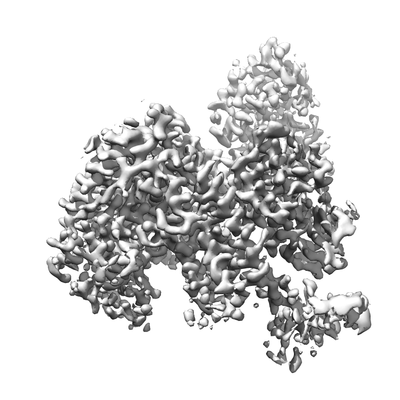 emd_0379.png emd_0379.png | 111.4 KB | ||
| Filedesc metadata | emd-0379.cif.gz | 7.3 KB | ||
| Archive directory |  http://ftp.pdbj.org/pub/emdb/structures/EMD-0379ftp://ftp.pdbj.org/pub/emdb/structures/EMD-0379 http://ftp.pdbj.org/pub/emdb/structures/EMD-0379ftp://ftp.pdbj.org/pub/emdb/structures/EMD-0379 | HTTPS FTP |
-Validation report
| Summary document | emd_0379_validation.pdf.gz | 359.7 KB | Display | EMDB validaton report |
|---|---|---|---|---|
| Full document | emd_0379_full_validation.pdf.gz | 359.3 KB | Display | |
| Data in XML | emd_0379_validation.xml.gz | 13.9 KB | Display | |
| Data in CIF | emd_0379_validation.cif.gz | 18.7 KB | Display | |
| Arichive directory | https://ftp.pdbj.org/pub/emdb/validation_reports/EMD-0379ftp://ftp.pdbj.org/pub/emdb/validation_reports/EMD-0379 | HTTPS FTP |
-Related structure data
| Related structure data | 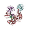 6n9uMC  0357C  0359C  0362C 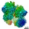 0363C 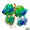 0364C 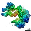 0365C 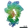 0380C 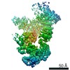 0381C  0382C 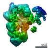 0386C 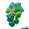 0387C 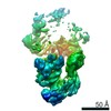 0388C 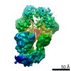 0389C 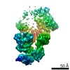 0390C 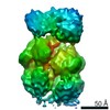 0391C 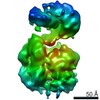 0392C 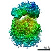 0393C 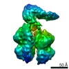 0394C 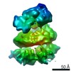 0395C  6n7iC  6n7nC 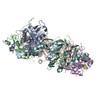 6n7sC 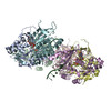 6n7tC 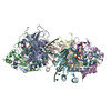 6n7vC 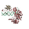 6n7wC 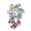 6n9vC 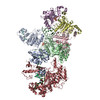 6n9wC 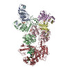 6n9xC C: citing same article ( M: atomic model generated by this map |
|---|---|
| Similar structure data |

-Links
| EMDB pages | EMDB (EBI/PDBe) / EMDataResource |
|---|---|
| Related items in Molecule of the Month |


-Map
| File | Download / File: emd_0379.map.gz / Format: CCP4 / Size: 244.1 MB / Type: IMAGE STORED AS FLOATING POINT NUMBER (4 BYTES) | ||||||||||||||||||||||||||||||||||||||||||||||||||||||||||||
|---|---|---|---|---|---|---|---|---|---|---|---|---|---|---|---|---|---|---|---|---|---|---|---|---|---|---|---|---|---|---|---|---|---|---|---|---|---|---|---|---|---|---|---|---|---|---|---|---|---|---|---|---|---|---|---|---|---|---|---|---|---|
| Annotation | Structure of gp5 DNA polymerase and two gp4 primase subunits complexed with trx, RNA/DNA hybrid, and incoming dTTP (from gp4-gp5 lagging-strand complex LagS1) | ||||||||||||||||||||||||||||||||||||||||||||||||||||||||||||
| Projections & slices | Image control
Images are generated by Spider. | ||||||||||||||||||||||||||||||||||||||||||||||||||||||||||||
| Voxel size | X=Y=Z: 0.86 Å | ||||||||||||||||||||||||||||||||||||||||||||||||||||||||||||
| Density |
| ||||||||||||||||||||||||||||||||||||||||||||||||||||||||||||
| Symmetry | Space group: 1 | ||||||||||||||||||||||||||||||||||||||||||||||||||||||||||||
| Details | EMDB XML:
CCP4 map header:
| ||||||||||||||||||||||||||||||||||||||||||||||||||||||||||||
 Z (Sec.)
 Y (Row.)
 X (Col.)


-Supplemental data
- Sample components
-Entire : gp5 DNA polymerase and two gp4 primase subunits complexed with tr...
| Entire | Name: gp5 DNA polymerase and two gp4 primase subunits complexed with trx, RNA/DNA hybrid, and incoming dTTP (from gp4-gp5 lagging-strand complex LagS1) |
|---|---|
| Components |
|
-Supramolecule #1: gp5 DNA polymerase and two gp4 primase subunits complexed with tr...
| Supramolecule | Name: gp5 DNA polymerase and two gp4 primase subunits complexed with trx, RNA/DNA hybrid, and incoming dTTP (from gp4-gp5 lagging-strand complex LagS1) type: complex / ID: 1 / Parent: 0 / Macromolecule list: #1-#4 |
|---|---|
| Source (natural) | Organism: Enterobacteria phage T7 (virus) |
-Macromolecule #1: DNA primase/helicase
| Macromolecule | Name: DNA primase/helicase / type: protein_or_peptide / ID: 1 / Number of copies: 2 / Enantiomer: LEVO EC number: Transferases; Transferring phosphorus-containing groups; Nucleotidyltransferases |
|---|---|
| Source (natural) | Organism: Enterobacteria phage T7 (virus) |
| Molecular weight | Theoretical: 62.73443 KDa |
| Recombinant expression | Organism:  |
| Sequence | String: MDNSHDSDSV FLYHIPCDNC GSSDGNSLFS DGHTFCYVCE KWTAGNEDTK ERASKRKPSG GKPMTYNVWN FGESNGRYSA LTARGISKE TCQKAGYWIA KVDGVMYQVA DYRDQNGNIV SQKVRDKDKN FKTTGSHKSD ALFGKHLWNG GKKIVVTEGE I DMLTVMEL ...String: MDNSHDSDSV FLYHIPCDNC GSSDGNSLFS DGHTFCYVCE KWTAGNEDTK ERASKRKPSG GKPMTYNVWN FGESNGRYSA LTARGISKE TCQKAGYWIA KVDGVMYQVA DYRDQNGNIV SQKVRDKDKN FKTTGSHKSD ALFGKHLWNG GKKIVVTEGE I DMLTVMEL QDCKYPVVSL GHGASAAKKT CAANYEYFDQ FEQIILMFDM DEAGRKAVEE AAQVLPAGKV RVAVLPCKDA NE CHLNGHD REIMEQVWNA GPWIPDGVVS ALSLRERIRE HLSSEESVGL LFSGCTGIND KTLGARGGEV IMVTSGSGMG KST FVRQQA LQWGTAMGKK VGLAMLQESV EETAEDLIGL HNRVRLRQSD SLKREIIENG KFDQWFDELF GNDTFHLYDS FAEA ETDRL LAKLAYMRSG LGCDVIILDH ISIVVSASGE SDERKMIDNL MTKLKGFAKS TGVVLVVICH LKNPDKGKAH EEGRP VSIT DLRGSGALRQ LSDTIIALER NQQGDMPNLV LVRILKCRFT GDTGIAGYME YNKETGWLEP SSYSGEEESH SESTDW SND TDF UniProtKB: DNA helicase/primase |
-Macromolecule #2: DNA-directed DNA polymerase
| Macromolecule | Name: DNA-directed DNA polymerase / type: protein_or_peptide / ID: 2 / Number of copies: 1 / Enantiomer: LEVO / EC number: DNA-directed DNA polymerase |
|---|---|
| Source (natural) | Organism: Enterobacteria phage T7 (virus) |
| Molecular weight | Theoretical: 79.805625 KDa |
| Recombinant expression | Organism: |
| Sequence | String: MIVSDIEANA LLESVTKFHC GVIYDYSTAE YVSYRPSDFG AYLDALEAEV ARGGLIVFHN GHKYDVPALT KLAKLQLNRE FHLPRENCI DTLVLSRLIH SNLKDTDMGL LRSGKLPGKR FGSHALEAWG YRLGEMKGEY KDDFKRMLEE QGEEYVDGME W WNFNEEMM ...String: MIVSDIEANA LLESVTKFHC GVIYDYSTAE YVSYRPSDFG AYLDALEAEV ARGGLIVFHN GHKYDVPALT KLAKLQLNRE FHLPRENCI DTLVLSRLIH SNLKDTDMGL LRSGKLPGKR FGSHALEAWG YRLGEMKGEY KDDFKRMLEE QGEEYVDGME W WNFNEEMM DYNVQDVVVT KALLEKLLSD KHYFPPEIDF TDVGYTTFWS ESLEAVDIEH RAAWLLAKQE RNGFPFDTKA IE ELYVELA ARRSELLRKL TETFGSWYQP KGGTEMFCHP RTGKPLPKYP RIKTPKVGGI FKKPKNKAQR EGREPCELDT REY VAGAPY TPVEHVVFNP SSRDHIQKKL QEAGWVPTKY TDKGAPVVDD EVLEGVRVDD PEKQAAIDLI KEYLMIQKRI GQSA EGDKA WLRYVAEDGK IHGSVNPNGA VTGRATHAFP NLAQIPGVRS PYGEQCRAAF GAEHHLDGIT GKPWVQAGID ASGLE LRCL AHFMARFDNG EYAHEILNGD IHTKNQIAAE LPTRDNAKTF IYGFLYGAGD EKIGQIVGAG KERGKELKKK FLENTP AIA ALRESIQQTL VESSQWVAGE QQVKWKRRWI KGLDGRKVHV RSPHAALNTL LQSAGALICK LWIIKTEEML VEKGLKH GW DGDFAYMAWV HDEIQVGCRT EEIAQVVIET AQEAMRWVGD HWNFRCLLDT EGKMGPNWAI CH UniProtKB: DNA-directed DNA polymerase |
-Macromolecule #3: RNA (5'-R(*AP*CP*CP*AP*G)-D(P*(DOC))-3')
| Macromolecule | Name: RNA (5'-R(*AP*CP*CP*AP*G)-D(P*(DOC))-3') / type: rna / ID: 3 / Number of copies: 1 |
|---|---|
| Source (natural) | Organism: Enterobacteria phage T7 (virus) |
| Molecular weight | Theoretical: 1.842206 KDa |
| Sequence | String: ACCAG(DOC) |
-Macromolecule #4: DNA (44-MER)
| Macromolecule | Name: DNA (44-MER) / type: dna / ID: 4 / Number of copies: 1 / Classification: DNA |
|---|---|
| Source (natural) | Organism: Enterobacteria phage T7 (virus) |
| Molecular weight | Theoretical: 13.402571 KDa |
| Sequence | String: (DT)(DT)(DT)(DT)(DT)(DA)(DG)(DC)(DT)(DG) (DG)(DT)(DC)(DA)(DT)(DT)(DT)(DT)(DT)(DT) (DT)(DT)(DT)(DT)(DT)(DT)(DT)(DT)(DT) (DT)(DT)(DT)(DT)(DT)(DT)(DT)(DT)(DT)(DT) (DT) (DT)(DT)(DT)(DT) |
-Macromolecule #5: ZINC ION
| Macromolecule | Name: ZINC ION / type: ligand / ID: 5 / Number of copies: 1 / Formula: ZN |
|---|---|
| Molecular weight | Theoretical: 65.409 Da |
-Macromolecule #6: THYMIDINE-5'-TRIPHOSPHATE
| Macromolecule | Name: THYMIDINE-5'-TRIPHOSPHATE / type: ligand / ID: 6 / Number of copies: 1 / Formula: TTP |
|---|---|
| Molecular weight | Theoretical: 482.168 Da |
| Chemical component information |  ChemComp-TTP: |
-Macromolecule #7: MAGNESIUM ION
| Macromolecule | Name: MAGNESIUM ION / type: ligand / ID: 7 / Number of copies: 2 / Formula: MG |
|---|---|
| Molecular weight | Theoretical: 24.305 Da |
-Experimental details
-Structure determination
| Method | cryo EM |
|---|---|
 Processing Processing | single particle reconstruction |
| Aggregation state | particle |
-Sample preparation
| Buffer | pH: 7.5 Component:
| ||||||||||||
|---|---|---|---|---|---|---|---|---|---|---|---|---|---|
| Grid | Details: unspecified | ||||||||||||
| Vitrification | Cryogen name: ETHANE / Chamber humidity: 100 % / Chamber temperature: 295 K / Instrument: FEI VITROBOT MARK I |
- Electron microscopy
| Microscope | FEI TITAN KRIOS |
|---|---|
| Image recording | Film or detector model: GATAN K2 SUMMIT (4k x 4k) / Detector mode: COUNTING / Average electron dose: 40.0 e/Å2 |
| Electron beam | Acceleration voltage: 300 kV / Electron source:  FIELD EMISSION GUN FIELD EMISSION GUN |
| Electron optics | Illumination mode: FLOOD BEAM / Imaging mode: BRIGHT FIELD |
| Experimental equipment |  Model: Titan Krios / Image courtesy: FEI Company |
+Image processing
-Atomic model buiding 1
| Initial model | PDB ID: Chain - Source name: PDB / Chain - Initial model type: experimental model |
|---|---|
| Refinement | Space: REAL / Protocol: FLEXIBLE FIT |
| Output model | PDB-6n9u: |
