 Movie
Movie Controller


+ Open data

- Basic information
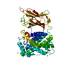
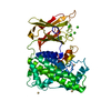
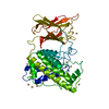
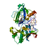
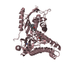
| Entry | Database: PDB / ID: 2wy6 | ||||||
|---|---|---|---|---|---|---|---|
| Title | Clostridium perfringens alpha-toxin strain NCTC8237 mutant T74I | ||||||
 Components Components | PHOSPHOLIPASE C | ||||||
 Keywords Keywords | HYDROLASE / CYTOLYSIS / HEMOLYSIS / MEMBRANE BINDING / VIRULENCE / GANGRENE DETERMINANT / C2 DOMAIN | ||||||
| Function / homology |  Function and homology information Function and homology informationphospholipase C / phosphatidylcholine phospholipase C activity / hemolysis in another organism / symbiont-mediated killing of host cell / toxin activity / hydrolase activity / extracellular region / zinc ion binding Similarity search - Function | ||||||
| Biological species |   CLOSTRIDIUM PERFRINGENS (bacteria) CLOSTRIDIUM PERFRINGENS (bacteria) | ||||||
| Method |  X-RAY DIFFRACTION / SYNCHROTRON / MOLECULAR REPLACEMENT / Resolution: 3.2 Å X-RAY DIFFRACTION / SYNCHROTRON / MOLECULAR REPLACEMENT / Resolution: 3.2 Å | ||||||
 Authors Authors | Vachieri, S.G. / Naylor, C.E. / Basak, A.K. | ||||||
 Citation Citation | Journal: Acta Crystallogr.,Sect.D / Year: 2011 Title: Comparison of a Nontoxic Variant of Clostridium Perfringens [Alpha]-Toxin with the Toxic Wild-Type Strain Authors: Vachieri, S.G. / Clark, G.C. / Alape-Giron, A. / Flores-Diaz, M. / Justin, N. / Naylor, C.E. / Titball, R.W. / Basak, A.K. #1: Journal: Nat.Struct.Biol. / Year: 1998Title: Structure of the Key Toxin in Gas Gangrene. Authors: Naylor, C.E. / Eaton, J.T. / Howells, A. / Justin, N. / Moss, D.S. / Titball, R.W. / Basak, A.K. #2: Journal: Microbiol.Immunol. / Year: 1996 Title: Threonine-74 is a Key Site for the Activity of Clostridium Perfringens Alpha-Toxin. Authors: Nagahama, M. / Sakurai, J. | ||||||
| History |
|
- Structure visualization
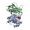
| Structure viewer | Molecule: MolmilJmol/JSmol |
|---|
- Downloads & links
-Download
| PDBx/mmCIF format | 2wy6.cif.gz | 225.7 KB | Display | PDBx/mmCIF format |
|---|---|---|---|---|
| PDB format | pdb2wy6.ent.gz | 178.6 KB | Display | PDB format |
| PDBx/mmJSON format | 2wy6.json.gz | Tree view | PDBx/mmJSON format | |
| Others |  Other downloads Other downloads |
-Validation report
| Arichive directory | https://data.pdbj.org/pub/pdb/validation_reports/wy/2wy6ftp://data.pdbj.org/pub/pdb/validation_reports/wy/2wy6 | HTTPS FTP |
|---|
-Related structure data
| Related structure data |  2wxtC  2wxuC  1ca1S C: citing same article ( S: Starting model for refinement |
|---|---|
| Similar structure data |


-Links
 PDBj
PDBj- Assembly
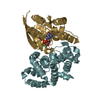
| Deposited unit | 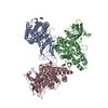
| ||||||||||||||||||||||||||||||||||||||||
|---|---|---|---|---|---|---|---|---|---|---|---|---|---|---|---|---|---|---|---|---|---|---|---|---|---|---|---|---|---|---|---|---|---|---|---|---|---|---|---|---|---|
| 1 | 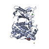
| ||||||||||||||||||||||||||||||||||||||||
| 2 | 
| ||||||||||||||||||||||||||||||||||||||||
| 3 | 
| ||||||||||||||||||||||||||||||||||||||||
| Unit cell |
| ||||||||||||||||||||||||||||||||||||||||
| Noncrystallographic symmetry (NCS) | NCS domain:
NCS domain segments:
NCS oper:
|
-Components
| #1: Protein | Mass: 42599.906 Da / Num. of mol.: 3 / Mutation: YES Source method: isolated from a genetically manipulated source Source: (gene. exp.) CLOSTRIDIUM PERFRINGENS (bacteria) / Strain: NCTC8237 / Plasmid: PT7BLUE / Production host: #2: Chemical |   Mass: 65.409 Da / Num. of mol.: 3 / Source method: obtained synthetically / Formula: Zn Mass: 65.409 Da / Num. of mol.: 3 / Source method: obtained synthetically / Formula: Zn#3: Chemical | ChemComp-CD /   Mass: 112.411 Da / Num. of mol.: 15 / Source method: obtained synthetically / Formula: Cd Mass: 112.411 Da / Num. of mol.: 15 / Source method: obtained synthetically / Formula: Cd#4: Chemical | ChemComp-CA /   Mass: 40.078 Da / Num. of mol.: 4 / Source method: obtained synthetically / Formula: Ca Mass: 40.078 Da / Num. of mol.: 4 / Source method: obtained synthetically / Formula: Ca#5: Chemical |   Mass: 92.094 Da / Num. of mol.: 2 / Source method: obtained synthetically / Formula: C3H8O3 Mass: 92.094 Da / Num. of mol.: 2 / Source method: obtained synthetically / Formula: C3H8O3Compound details | ENGINEERED RESIDUE IN CHAIN A, THR 102 TO ILE ENGINEERED RESIDUE IN CHAIN B, THR 102 TO ILE ...ENGINEERED | |
|---|
-Experimental details
-Experiment
| Experiment | Method: X-RAY DIFFRACTION / Number of used crystals: 1 |
|---|
- Sample preparation
| Crystal | Density Matthews: 2.6 Å3/Da / Density % sol: 52.6 % / Description: NONE |
|---|---|
| Crystal grow | pH: 7.6 Details: 1 M NA ACETATE, 0,1 M NA-HEPES, PH 7.6, 0.05 M CDSO4 |
-Data collection
| Diffraction | Mean temperature: 100 K |
|---|---|
| Diffraction source | Source: SYNCHROTRON / Site: ESRF  / Beamline: ID14-2 / Wavelength: 0.933 / Beamline: ID14-2 / Wavelength: 0.933 |
| Detector | Type: MARRESEARCH / Detector: CCD / Date: Dec 11, 2003 / Details: MIRRORS |
| Radiation | Monochromator: SILICON (311) / Protocol: SINGLE WAVELENGTH / Monochromatic (M) / Laue (L): M / Scattering type: x-ray |
| Radiation wavelength | Wavelength: 0.933 Å / Relative weight: 1 |
| Reflection | Resolution: 3.2→29.74 Å / Num. obs: 22436 / % possible obs: 99.8 % / Observed criterion σ(I): 0 / Redundancy: 7.8 % / Biso Wilson estimate: 88.88 Å2 / Rmerge(I) obs: 0.09 / Net I/σ(I): 22.4 |
| Reflection shell | Resolution: 3.2→3.4 Å / Redundancy: 8 % / Rmerge(I) obs: 0.54 / Mean I/σ(I) obs: 3.5 / % possible all: 100 |
- Processing
| Software |
| |||||||||||||||||||||||||||||||||||||||||||||||||||||||||||||||
|---|---|---|---|---|---|---|---|---|---|---|---|---|---|---|---|---|---|---|---|---|---|---|---|---|---|---|---|---|---|---|---|---|---|---|---|---|---|---|---|---|---|---|---|---|---|---|---|---|---|---|---|---|---|---|---|---|---|---|---|---|---|---|---|---|
| Refinement | Method to determine structure: MOLECULAR REPLACEMENT Starting model: PDB ENTRY 1CA1 WITHOUT RESIDUES 75-84 Resolution: 3.2→24.914 Å / SU ML: 0.41 / σ(F): 1.39 / Phase error: 27.32 / Stereochemistry target values: ML
| |||||||||||||||||||||||||||||||||||||||||||||||||||||||||||||||
| Solvent computation | Shrinkage radii: 0.9 Å / VDW probe radii: 1.11 Å / Solvent model: FLAT BULK SOLVENT MODEL / Bsol: 50.698 Å2 / ksol: 0.306 e/Å3 | |||||||||||||||||||||||||||||||||||||||||||||||||||||||||||||||
| Displacement parameters | Biso mean: 92.37 Å2
| |||||||||||||||||||||||||||||||||||||||||||||||||||||||||||||||
| Refinement step | Cycle: LAST / Resolution: 3.2→24.914 Å
| |||||||||||||||||||||||||||||||||||||||||||||||||||||||||||||||
| Refine LS restraints |
| |||||||||||||||||||||||||||||||||||||||||||||||||||||||||||||||
| Refine LS restraints NCS |
| |||||||||||||||||||||||||||||||||||||||||||||||||||||||||||||||
| LS refinement shell |
|
