+ Open data
- Basic information
| Entry | Database: PDB / ID: 2f9g | ||||||
|---|---|---|---|---|---|---|---|
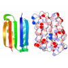
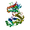
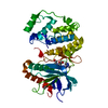
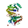
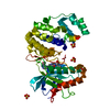
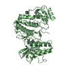
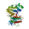
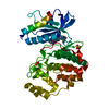
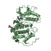
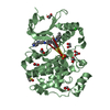
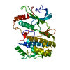
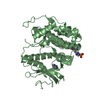
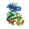
| Title | Crystal structure of Fus3 phosphorylated on Tyr182 | ||||||
 Components Components | Mitogen-activated protein kinase FUS3 | ||||||
 Keywords Keywords | TRANSFERASE / MAP kinase | ||||||
| Function / homology |  Function and homology information Function and homology information: / phospho-PLA2 pathway / : / : / ERK/MAPK targets / Signalling to ERK5 / ERKs are inactivated / Senescence-Associated Secretory Phenotype (SASP) / : / Ca2+ pathway ...: / phospho-PLA2 pathway / : / : / ERK/MAPK targets / Signalling to ERK5 / ERKs are inactivated / Senescence-Associated Secretory Phenotype (SASP) / : / Ca2+ pathway / RAF/MAP kinase cascade / : / Gastrin-CREB signalling pathway via PKC and MAPK / Estrogen-stimulated signaling through PRKCZ / : / Signal transduction by L1 / : / : / pheromone response MAPK cascade / Recycling pathway of L1 / response to pheromone triggering conjugation with cellular fusion / pheromone-dependent signal transduction involved in conjugation with cellular fusion / invasive growth in response to glucose limitation / Activation of the AP-1 family of transcription factors / Regulation of HSF1-mediated heat shock response / Transcriptional and post-translational regulation of MITF-M expression and activity / transposable element silencing / mating projection tip / MAPK6/MAPK4 signaling / MAP kinase activity / mitogen-activated protein kinase / negative regulation of MAPK cascade / Neutrophil degranulation / positive regulation of protein export from nucleus / cytoplasmic stress granule / periplasmic space / protein kinase activity / intracellular signal transduction / cell division / protein serine kinase activity / protein serine/threonine kinase activity / mitochondrion / ATP binding / identical protein binding / nucleus / cytoplasm Similarity search - Function | ||||||
| Biological species |  | ||||||
| Method |  X-RAY DIFFRACTION / X-RAY DIFFRACTION /  SYNCHROTRON / SYNCHROTRON /  MOLECULAR REPLACEMENT / Resolution: 2.1 Å MOLECULAR REPLACEMENT / Resolution: 2.1 Å | ||||||
 Authors Authors | Bhattacharyya, R.P. / Remenyi, A. / Good, M.C. / Bashor, C.J. / Falick, A.M. / Lim, W.A. | ||||||
 Citation Citation |  Journal: Science / Year: 2006 Journal: Science / Year: 2006Title: The Ste5 scaffold allosterically modulates signaling output of the yeast mating pathway. Authors: Bhattacharyya, R.P. / Remenyi, A. / Good, M.C. / Bashor, C.J. / Falick, A.M. / Lim, W.A. | ||||||
| History |
| ||||||
| Remark 400 | COMPOUND ONE OF THE TWO RESIDUES IN THE ACTIVATION LOOP (TYR182) IS PHOSPHORYLATED. (NOTE: FOR MAP ...COMPOUND ONE OF THE TWO RESIDUES IN THE ACTIVATION LOOP (TYR182) IS PHOSPHORYLATED. (NOTE: FOR MAP KINASES PHOSPHORYLATION OF BOTH RESIDUES (THR180 AND TYR182) IS REQUIRED FOR FULL ACTIVATION.) TYR182 PHOSPHORYLATION RENDERS THE FUS3 ACTIVATION LOOP MORE FLEXIBLE. THE PHOSPHORYLATED RESIDUE CAN NOT BE SEEN IN THE ELECTRON DENSITY. |
- Structure visualization
| Structure viewer | Molecule:  Molmil Molmil Jmol/JSmol Jmol/JSmol |
|---|
- Downloads & links
- Download
| PDBx/mmCIF format |  2f9g.cif.gz 2f9g.cif.gz | 84 KB | Display |  PDBx/mmCIF format PDBx/mmCIF format |
|---|---|---|---|---|
| PDB format |  pdb2f9g.ent.gz pdb2f9g.ent.gz | 62.1 KB | Display |  PDB format PDB format |
| PDBx/mmJSON format |  2f9g.json.gz 2f9g.json.gz | Tree view |  PDBx/mmJSON format PDBx/mmJSON format | |
| Others |  Other downloads Other downloads |
-Validation report
| Summary document |  2f9g_validation.pdf.gz 2f9g_validation.pdf.gz | 754.9 KB | Display |  wwPDB validaton report wwPDB validaton report |
|---|---|---|---|---|
| Full document |  2f9g_full_validation.pdf.gz 2f9g_full_validation.pdf.gz | 763.7 KB | Display | |
| Data in XML |  2f9g_validation.xml.gz 2f9g_validation.xml.gz | 16.2 KB | Display | |
| Data in CIF |  2f9g_validation.cif.gz 2f9g_validation.cif.gz | 22.3 KB | Display | |
| Arichive directory |  https://data.pdbj.org/pub/pdb/validation_reports/f9/2f9g https://data.pdbj.org/pub/pdb/validation_reports/f9/2f9g ftp://data.pdbj.org/pub/pdb/validation_reports/f9/2f9g ftp://data.pdbj.org/pub/pdb/validation_reports/f9/2f9g | HTTPS FTP |
-Related structure data
| Related structure data |  2f49C  2fa2C  2b9fS S: Starting model for refinement C: citing same article ( |
|---|---|
| Similar structure data |
- Links
Links
- Assembly
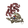
| Deposited unit | 
| ||||||||
|---|---|---|---|---|---|---|---|---|---|
| 1 |
| ||||||||
| Unit cell |
|
- Components
| #1: Protein | Mass: 40906.996 Da / Num. of mol.: 1 Source method: isolated from a genetically manipulated source Source: (gene. exp.)  Description: Recombinant Fus3 was incubated with an activator peptide from Ste5 to achieve full phosphorylation on Tyr182 by autophsophorylation. Gene: FUS3, DAC2 / Plasmid: pBH4-Fus3 / Production host:  |
|---|---|
| #2: Chemical | ChemComp-MG / |
| #3: Chemical | ChemComp-ADP / |
| #4: Water | ChemComp-HOH / |
| Has protein modification | N |
-Experimental details
-Experiment
| Experiment | Method:  X-RAY DIFFRACTION / Number of used crystals: 1 X-RAY DIFFRACTION / Number of used crystals: 1 |
|---|
- Sample preparation
| Crystal | Density Matthews: 1.87 Å3/Da / Density % sol: 34.09 % |
|---|---|
| Crystal grow | Temperature: 293 K / Method: vapor diffusion, hanging drop / pH: 6.1 Details: 25-28% PEG1000, 0.1M MES, 5-10% MPD, pH 6.1, VAPOR DIFFUSION, HANGING DROP, temperature 293K |
-Data collection
| Diffraction | Mean temperature: 100 K |
|---|---|
| Diffraction source | Source:  SYNCHROTRON / Site: SYNCHROTRON / Site:  ALS ALS  / Beamline: 8.3.1 / Wavelength: 1.115889 Å / Beamline: 8.3.1 / Wavelength: 1.115889 Å |
| Detector | Type: ADSC QUANTUM 4 / Detector: CCD / Date: Apr 23, 2004 |
| Radiation | Monochromator: Double crystal / Protocol: SINGLE WAVELENGTH / Monochromatic (M) / Laue (L): M / Scattering type: x-ray |
| Radiation wavelength | Wavelength: 1.115889 Å / Relative weight: 1 |
| Reflection | Resolution: 2.1→50 Å / Num. all: 18096 / Num. obs: 18096 / % possible obs: 97.3 % / Observed criterion σ(F): 0 / Observed criterion σ(I): 0 / Redundancy: 3 % / Rsym value: 0.09 / Net I/σ(I): 10.7 |
| Reflection shell | Resolution: 2.1→2.18 Å / Mean I/σ(I) obs: 2.2 / Num. unique all: 1574 / Rsym value: 0.346 / % possible all: 86.6 |
- Processing
| Software |
| |||||||||||||||||||||||||
|---|---|---|---|---|---|---|---|---|---|---|---|---|---|---|---|---|---|---|---|---|---|---|---|---|---|---|
| Refinement | Method to determine structure:  MOLECULAR REPLACEMENT MOLECULAR REPLACEMENTStarting model: PDB entry 2B9F Resolution: 2.1→20 Å / Cross valid method: THROUGHOUT / σ(F): 0 / Stereochemistry target values: Engh & Huber
| |||||||||||||||||||||||||
| Refinement step | Cycle: LAST / Resolution: 2.1→20 Å
| |||||||||||||||||||||||||
| Refine LS restraints |
|
 Movie
Movie Controller



 PDBj
PDBj
