 Movie
Movie Controller


+ Open data

- Basic information
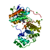
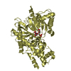
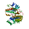
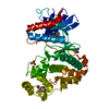
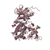
| Entry | Database: PDB / ID: 2f49 | ||||||
|---|---|---|---|---|---|---|---|
| Title | Crystal structure of Fus3 in complex with a Ste5 peptide | ||||||
 Components Components |
| ||||||
 Keywords Keywords | TRANSFERASE / protein-petide complex | ||||||
| Function / homology |  Function and homology information Function and homology information: / phospho-PLA2 pathway / : / : / ERK/MAPK targets / Signalling to ERK5 / Senescence-Associated Secretory Phenotype (SASP) / : / Ca2+ pathway / RAF/MAP kinase cascade ...: / phospho-PLA2 pathway / : / : / ERK/MAPK targets / Signalling to ERK5 / Senescence-Associated Secretory Phenotype (SASP) / : / Ca2+ pathway / RAF/MAP kinase cascade / : / Gastrin-CREB signalling pathway via PKC and MAPK / Estrogen-stimulated signaling through PRKCZ / : / : / : / ERKs are inactivated / pheromone response MAPK cascade / Signal transduction by L1 / Recycling pathway of L1 / response to pheromone triggering conjugation with cellular fusion / : / invasive growth in response to glucose limitation / Regulation of HSF1-mediated heat shock response / Activation of the AP-1 family of transcription factors / Transcriptional and post-translational regulation of MITF-M expression and activity / transposable element silencing / mating projection tip / MAPK6/MAPK4 signaling / MAP kinase activity / mitogen-activated protein kinase / negative regulation of MAPK cascade / Neutrophil degranulation / positive regulation of protein export from nucleus / cytoplasmic stress granule / protein kinase activity / periplasmic space / intracellular signal transduction / protein serine kinase activity / cell division / protein serine/threonine kinase activity / mitochondrion / ATP binding / identical protein binding / nucleus / cytoplasm Similarity search - Function | ||||||
| Biological species |  | ||||||
| Method |  X-RAY DIFFRACTION / SYNCHROTRON / MOLECULAR REPLACEMENT / Resolution: 1.9 Å X-RAY DIFFRACTION / SYNCHROTRON / MOLECULAR REPLACEMENT / Resolution: 1.9 Å | ||||||
 Authors Authors | Remenyi, A. | ||||||
 Citation Citation | Journal: Science / Year: 2006 Title: The Ste5 scaffold allosterically modulates signaling output of the yeast mating pathway Authors: Bhattacharyya, R.P. / Remenyi, A. / Good, M.C. / Bashor, C.J. / Falick, A.M. / Lim, W.A. | ||||||
| History |
|
- Structure visualization
| Structure viewer | Molecule: MolmilJmol/JSmol |
|---|
- Downloads & links
-Download
| PDBx/mmCIF format | 2f49.cif.gz | 161.2 KB | Display | PDBx/mmCIF format |
|---|---|---|---|---|
| PDB format | pdb2f49.ent.gz | 125.4 KB | Display | PDB format |
| PDBx/mmJSON format | 2f49.json.gz | Tree view | PDBx/mmJSON format | |
| Others |  Other downloads Other downloads |
-Validation report
| Arichive directory | https://data.pdbj.org/pub/pdb/validation_reports/f4/2f49ftp://data.pdbj.org/pub/pdb/validation_reports/f4/2f49 | HTTPS FTP |
|---|
-Related structure data
| Related structure data |  2f9gC  2fa2C  2b9fS S: Starting model for refinement C: citing same article ( |
|---|---|
| Similar structure data |


-Links
 PDBj
PDBj


- Assembly
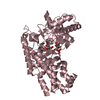
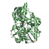
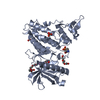
| Deposited unit | 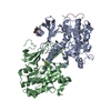
| ||||||||
|---|---|---|---|---|---|---|---|---|---|
| 1 | 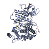
| ||||||||
| 2 | 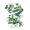
| ||||||||
| 3 |
| ||||||||
| 4 | 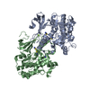
| ||||||||
| 5 | 
| ||||||||
| Unit cell |
| ||||||||
| Details | The biological unit is a protein-peptide complex between chain A and C |
-Components
| #1: Protein | Mass: 40809.047 Da / Num. of mol.: 2 / Fragment: Fus3 / Mutation: T180V, Y182F Source method: isolated from a genetically manipulated source Source: (gene. exp.) Gene: FUS3, DAC2 / Plasmid: pBH4-Fus3 / Production host:  #2: Protein/peptide | | Mass: 3349.769 Da / Num. of mol.: 1 / Fragment: Fus3 binding region of STE5 / Source method: obtained synthetically Details: This peptide was chemically synthesized but this sequence naturally occurs in the Ste5 protein from S. cerevisiae #3: Chemical |   Mass: 24.305 Da / Num. of mol.: 2 / Source method: obtained synthetically / Formula: Mg Mass: 24.305 Da / Num. of mol.: 2 / Source method: obtained synthetically / Formula: Mg#4: Chemical | ChemComp-SCN /   Mass: 58.082 Da / Num. of mol.: 7 / Source method: obtained synthetically / Formula: CNS Mass: 58.082 Da / Num. of mol.: 7 / Source method: obtained synthetically / Formula: CNS#5: Water | ChemComp-HOH / |  Mass: 18.015 Da / Num. of mol.: 478 / Source method: isolated from a natural source / Formula: H2O Mass: 18.015 Da / Num. of mol.: 478 / Source method: isolated from a natural source / Formula: H2O |
|---|
-Experimental details
-Experiment
| Experiment | Method: X-RAY DIFFRACTION / Number of used crystals: 1 |
|---|
- Sample preparation
| Crystal | Density Matthews: 2.69 Å3/Da / Density % sol: 54.33 % |
|---|---|
| Crystal grow | Temperature: 293 K / Method: vapor diffusion / pH: 6.1 Details: 18% PEG3350, 0.1M Mes, 10% MPD, 0.2M KSCN, pH 6.1, VAPOR DIFFUSION, temperature 293K |
-Data collection
| Diffraction | Mean temperature: 100 K |
|---|---|
| Diffraction source | Source: SYNCHROTRON / Site: ALS  / Beamline: 8.3.1 / Wavelength: 1.1159 Å / Beamline: 8.3.1 / Wavelength: 1.1159 Å |
| Detector | Type: ADSC QUANTUM 4 / Detector: CCD / Date: Mar 13, 2004 |
| Radiation | Monochromator: double crystal / Protocol: SINGLE WAVELENGTH / Monochromatic (M) / Laue (L): M / Scattering type: x-ray |
| Radiation wavelength | Wavelength: 1.1159 Å / Relative weight: 1 |
| Reflection | Resolution: 1.9→50 Å / Num. all: 69388 / Num. obs: 69388 / % possible obs: 95.3 % / Observed criterion σ(F): 0 / Observed criterion σ(I): 0 / Redundancy: 4.4 % / Rsym value: 0.07 / Net I/σ(I): 15.3 |
| Reflection shell | Resolution: 1.9→1.97 Å / Mean I/σ(I) obs: 2.4 / Num. unique all: 5920 / Rsym value: 0.5 / % possible all: 83.2 |
- Processing
| Software |
| |||||||||||||||||||||||||
|---|---|---|---|---|---|---|---|---|---|---|---|---|---|---|---|---|---|---|---|---|---|---|---|---|---|---|
| Refinement | Method to determine structure: MOLECULAR REPLACEMENT Starting model: PDB entry 2B9F Resolution: 1.9→20 Å / σ(F): 0 / Stereochemistry target values: Engh & Huber
| |||||||||||||||||||||||||
| Refinement step | Cycle: LAST / Resolution: 1.9→20 Å
| |||||||||||||||||||||||||
| Refine LS restraints |
|
